NICE on Lyme disease this month – just in time for the weather to pick up and the tics to start biting. Also a reminder on the risk factors for SIDS, what to do in a terrorist attack, how to manage a child with a non-blanching rash and a discussion on the use of the antistreptolysin O titre. Do leave comments below:
Late night musings on ASOT
A patient was referred to me in the paediatric cardiology clinic because of a risk that he may have had missed Kawasaki’s disease a couple of weeks earlier and was therefore at risk of having coronary artery aneurysms. The referring doctor had carried out an antistreptolysin O titer (ASOT) in case the symptoms of a red, sore mouth, rash and later peeling fingers had been secondary to a streptococcal infection rather than KD. The result came back as 400units/ml (normal is < 200units/ml). The child was very well when I saw him and had a normal echocardiogram. What should I do with the elevated ASOT result?
I needed a quick text box as a gap filler for the April edition of the Paediatric Pearls newsletter and thought ASOT results would be a suitable topic but, when I sat down to write it, I opened up a can of worms. No one really knows what to do with high ASOTs in a well child. In fact, authors can’t even agree on whether 400 is elevated in a young person.
My reading list is at the foot of this article. Salient points from these sources are summarised below.
- The ASOT is ordered primarily to determine whether a previous group A Streptococcus infection has caused a poststreptococcal complication, such as rheumatic fever or glomerulonephritis. So the start point should be on-going clinical symptoms of strep infection or the effect of a recent infection. If used in this way, it can be a useful pointer to a causative organism and will guide management. Rheumatic fever is treated with long term antibiotics. The ASO test does not predict whether complications will occur following a strep infection, nor does it predict the type or severity of the disease. If symptoms of rheumatic fever or glomerulonephritis are present, an elevated ASO level may be used to help confirm the diagnosis.
- ASO antibodies are produced a week to a month after an initial strep infection. The amount of ASO antibody (titer) peaks at 3 to 5 weeks after the illness and then tapers off but may remain detectable for several months after the strep infection has resolved.
- A negative ASO or ASO that is present at very low titers means the person tested most likely has not had a recent strep infection. This is especially true if a sample taken 10 to 14 days later is also negative (low titer of antibody) and if an anti-DNase B test is also negative (low titer of antibody). A small percentage of people with a complication related to a strep infection will not have an elevated ASO. This is especially true with glomerulonephritis that may develop after a skin strep infection.
- An elevated titer of antibody (positive ASO) or an ASO titer that is rising means that it is likely that the person tested has had a recent strep infection. ASO titers that are initially high and then decline suggest that an infection has occurred and may be resolving.
My conclusion at the end of reading about ASOT and the management of streptococcal infections and complications is that I should only do the ASOT if the child is symptomatic. If I think they have rheumatic fever, I should treat with antibiotics for a long time (up to 10 years in some cases). If they do not satisfy the Jones criteria for rheumatic fever and indeed are well now, I do not need to blindly treat an elevated ASOT but it may be prudent to repeat the test a couple of weeks later to ensure it is dropping.
Very good summary article on rheumatic fever: https://patient.info/doctor/rheumatic-fever-pro
Why treat sore throats at all? https://www.ncbi.nlm.nih.gov/pmc/articles/PMC1949249/
Cochrane on short term antibiotics: https://www.ncbi.nlm.nih.gov/pubmed/22895944
https://www.annemergmed.com/article/S0196-0644(13)01448-0/fulltext on same topic
https://www.uptodate.com/contents/treatment-and-prevention-of-streptococcal-pharyngitis
March 2018 PDF in time for Easter
NICE on faltering growth this month, paediatric stroke, a reminder of the new epilepsy classification and a contribution from the safeguarding team on what constitutes a “legal high”? Do leave comments below:
Epilepsy classification changes again…
Actually the classification of seizures changed in July 2017 but I’ve only just been brought up to date by Emily O’Connor, a medical student who writes blog posts for Paediatric Pearls. Here is her article:
In 2017 the International League Against Epilepsy revised their classification of seizure types, with the aim of creating greater flexibility, accuracy and transparency in the naming of seizures. Below, is a brief guide to applying this new approach to classification and a summary of the changes in terminology.
The new approach can be applied by asking two or three questions about the seizure:
- Where was the onset of the seizure?
- It could be: focal/generalised/focal to bilateral/unknown
- What was the patient’s level of awareness during the seizure? – FOR FOCAL SEIZURES ONLY
- It could be: focal aware/focal impaired awareness
- What was the first prominent sign or symptom of the seizure?
- It could be: motor/non-motor
- This can then be further classified according to the specific symptom
This new classification system for seizures has led to a change in some of the traditional terminology used to describe seizure types, the below table shows a summary of these changes:
| Traditional/‘Obsolete’ Term | New/‘Replacement’ Term |
| Partial seizure | Focal seizure |
| Simple partial seizure | Focal aware seizure |
| Complex partial/Dyscognitive seizure | Focal impaired awareness seizure |
| Psychic seizure | Cognitive seizure |
| Primary generalised seizure | Generalised seizure |
| Secondary generalised seizure | Focal to bilateral tonic-clonic seizure |
For more information on the ILEA 2017 classification system, please see the below references:
1. Fisher et al. Operational classification of seizure types by the International League Against Epilepsy: Position Paper of the ILAE Commission for Classification and Terminology. Epilepsia. 2017. 58. 4. 522-530.
2. Epilepsy Foundation of America. 2017 Revised Classification of Seizures. [online] Epilepsy Foundation of America. 2017. 18/02/2018. <https://www.epilepsy.com/article/2016/12/2017-revised-classification-seizures>
February’s newsletter 2018
What constitutes sexualised behaviour in a 4 year old? This and the childhood asthma control test, this month, toddler fractures and the PCV vaccine. Do leave comments below.
Journal Club: Lumbar Punctures in Children with Febrile Seizures
Journal Club is a revamped monthly feature in the Paediatric Pearls newsletter. I’m happy to receive submissions from any primary or secondary care journal club you are running as long as the paper is relevant to front line health professionals working with children. Please contact me through the contact page.
With thanks this week to Dr Saskia Wills who took us through a paper on the need (or not) for LPs in children with complex febrile seizures. Her full presentation is here.
In brief:
- The definition of a febrile seizure in this paper is a seizure in a child 6 months to 5 years with a fever >38o and without an underlying CNS infection or a history of afebrile seizures
- They occur in 2-4% of children <5yrs (peak at 12-18 months)
- They are classified as complex if they last >15 minutes, have a focal onset, or there are multiple episodes within 24 hours
- They are often associated with viral infections, especially HHV6
- The risk is slightly higher in boys and those with a family history of febrile convulsion
- 1/3 of children will have another febrile seizure in the future, but very few (2.4%) go on to have epilepsy. (The risk of epilepsy, which varies with different presenting features, is discussed here)
- In a retrospective French study of otherwise well children presenting with complex febrile seizures, only 5 out of 839 (0.7%) had confirmed bacterial meningitis. All of these had had a prolonged seizure plus some ongoing abnormal neurology or sign suggestive of CNS infection. The study concluded that in children with complex febrile seizures but no other signs of CNS infection, LP usually isn’t necessary. The risk of proven CNS disease is higher in those under 1yr and with a prolonged seizure. This study didn’t look at children who had other risk factors for meningitis, such as immunodeficiency.
Paper studied: Guedj R1, Chappuy H2 et al. Do All Children Who Present With a Complex Febrile Seizure Need a Lumbar Puncture? Ann Emerg Med. 2017 Jul;70(1):52-62. PubMed Link.
Managing a Child with a Decreased Conscious Level
With thanks to Dr Dilshad Marikar for looking at the 2016 RCPCH material on managing a child with a decreased conscious level, prompted by his being on call when a 14 year old was brought to the ED with a GCS of 3.
The RCPCH algorithm has a child between 4 weeks and 18 years old enter the Decreased Consciousness (DeCon) pathway if the AVPU is “P” or “U” or if there is a new finding of GCS ≤ 14 which seems quite a low threshold and means that we will all need to use this guideline at some point. See: https://www.rcpch.ac.uk/system/files/protected/page/RCPCH%20DeCon%20Poster%20.pdf – incorporates how to identify and manage the situation and differential diagnoses. The full guideline is available here and the recommendations summary PDF, here. Some salient points:
- Consider intubation if GCS < 8 and child not improving
- Give oxygen if O2 saturation is ≤ 95%
- Check capillary blood glucose within 15 minutes
- Don’t overlook the possibility of NAI or safeguarding issues
Who Has Not Been Asked About Nappy Rash?
The Avon Longitudinal Study of Parents and Children (ALSPAC) study collected information about nappy rash using self-completed questionnaires answered by parents at the end of the first four weeks of their baby’s life. The study found that 25% of the babies had experienced napkin dermatitis.
NICE has a comprehensive clinical knowledge summary on nappy rash here. Salient points:
- Skin swabs are not recommended for the management of nappy rash as the results are difficult to interpret.
- Both Candida and bacteria (such as Staphylococcus aureus) colonize healthy skin and a skin swab may be positive when infection is not present.
- A swab should only be taken when a secondary bacterial infection is suspected, to guide choice of antibiotic
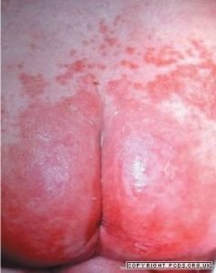
This picture and more available on the excellent Primary Care Dermatology Society website. Compare this candidiasis picture with ammoniacal dermatitis and napkin eczema.
- Consider using nappies with the greatest absorbency (for example, disposable gel matrix nappies)
- Leave nappies off for as long as is practically possible. Clean and change the child as soon as possible after wetting or soiling. Use water, or fragrance-free and alcohol-free baby wipes.
- Dry gently after cleaning — avoid vigorous rubbing.
- Bath the child daily — but avoid excessive bathing (such as more than twice a day) which may dry the skin.
- Do not use soap, bubble bath, or lotions. Advise about skin care.
- Prescribe a barrier preparation to apply thinly at each nappy change, to protect the skin. Zinc and Castor Oil ointment BP or Metanium® ointment are recommended. Alternatively, white soft paraffin BP ointment or dexpanthenol 5% ointment (Bepanthen®) could be used.
- For children over 1 month of age, consider prescribing topical hydrocortisone 0.5% or 1% cream once a day for 7 days max.
Signs of Raised Intracranial Pressure (ICP)
From APLS manual 6E
- Abnormal oculocephalic reflexes (avoid in patients with neck injuries):
When the head is turned to the left or right a normal response is for the eyes to move away from the head movement; an abnormal response is no (or random) movement. See video for a demo of normal reflexes. - Abnormal Posture:
Decorticate (flexed arms, extended legs)
Decerebrate (extended arms, extended legs)
Posturing may need to be elicited by a painful stimulus - Abnormal pupillary responses: unilateral or bilateral dilatation suggests raised ICP
- Abnormal breathing patterns: There are several recognisable breathing pattern abnormalities in raised ICP. However they are often changeable and may vary from hyperventilation to Cheyne-Stokes breathing to apnoea
- Cushing’s Triad: Hypertension, Bradycardia and breathing pattern abnormalities are a late sign of raised ICP
Winter, Vitamin D and Rickets
It’s dark and sun-less again in the UK and everyone’s Vitamin D levels will be at rock bottom over the next couple of months. Rickets is not rare in London and neither are consequent hypocalcaemic fits in our babies and teenagers unfortunately. Hackney CCG has an easy to follow algorithm for prevention and management of Vitamin D deficiency: you can find it here.
There’s even a table which tells you which vitamin preparations are suitable for vegetarians or vegans, which are Kosher and Halal certified and which to avoid in peanut allergy.