I was a bit overloaded in December and couldn’t manage to get a newsletter out. The nurses in my own ED noticed but I don’t think anyone else was particularly bothered! Hope the Christmas break went well for everyone. January 2013 brings some more on speech development, a reminder of the BTS 2008 guideline on cough, another plug for vitamin supplementation and part 2 of Jess Spedding’s minor injuries series. Do leave comments below.
Category Archives: For General Practitioners
November 2012 published!
The common assessment framework triangle for assessing children in need this month with some tips on how to press the right buttons with children’s social care referrals. Also a bit on stabilisation and transfer for the ED teams, a reminder not to use 0.18% saline and the start of a minor injuries series. Talipes for the GPs and paediatricians among you.
Talipes equinovarus
Talipes (Neonatal Clubfoot) with thanks to Dr Mujahid Hasan and the paediatric physiotherapy department at Barts Health
Newborn babies can present with one of two types of Talipes:
- Congenital Talipes Equinovarus (CTEV or fixed/structural Talipes)
- Positional foot problems
Click here for the Whipps Cross physiotherapists and Muj’s complete, illustrated article.
NICE headaches
- worsening headache with fever
- sudden-onset headache reaching maximum intensity within 5 minutes
- new-onset neurological deficit
- new-onset cognitive dysfunction
- change in personality
- impaired level of consciousness
- recent (typically within the past 3 months) head trauma
- headache triggered by cough, valsalva (trying to breathe out with nose and mouth blocked) or sneeze
- headache triggered by exercise
- orthostatic headache (headache that changes with posture)
- symptoms suggestive of giant cell arteritis
- symptoms and signs of acute narrow-angle glaucoma
- a substantial change in the characteristics of their headache.
- compromised immunity, caused, for example, by HIV or immunosuppressive drugs
- age under 20 years and a history of malignancy
- a history of malignancy known to metastasise to the brain
- vomiting without other obvious cause.
- frequency, duration and severity of headaches
- any associated symptoms
- all prescribed and over the counter medications taken to relieve headaches
- possible precipitants
- relationship of headaches to menstruation.
| Headache feature | Tension-type headache | Migraine (with or without aura) | Cluster headache | |||
| Pain location1 | Bilateral | Unilateral or bilateral | Unilateral (around the eye, above the eye and along the side of the head/face) |
|||
| Pain quality | Pressing/tightening (non-pulsating) | Pulsating (throbbing or banging in young people aged 12–17 years) | Variable (can be sharp, boring, burning, throbbing or tightening) | |||
| Pain intensity | Mild or moderate | Moderate or severe | Severe or very severe | |||
| Effect on activities | Not aggravated by routine activities of daily living | Aggravated by, or causes avoidance of, routine activities of daily living | Restlessness or agitation | |||
| Other symptoms | None | Unusual sensitivity to light and/or sound or nausea and/or vomiting Aura2 Symptoms can occur with or without headache and:
Typical aura symptoms include visual symptoms such as flickering lights,
spots or lines and/or partial loss of vision; sensory symptoms such as numbness and/or pins and needles; and/or speech disturbance. |
On the same side as the headache:
|
|||
| Duration of headache | 30 minutes–continuous | 4–72 hours in adults 1–72 hours in young people aged 12–17 years |
15–180 minutes | |||
| Frequency of headache | < 15 days per month | ≥ 15 days per month for more than 3 months | < 15 days per month | ≥ 15 days per month for more than 3 months | 1 every other day to 8 per day3, with remission4 > 1 month |
1 every other day to 8 per day3,
with a continuous remission4 <1 month
in a
12-month period
|
| Diagnosis | Episodic
tension-type headache
|
Chronic tension-type headache5 | Episodic migraine (with or without aura) | Chronic migraine6 (with or without aura) | Episodic cluster headache | Chronic cluster headache |
| 1 Headache pain can be felt in the head, face or neck. 2 See recommendations 1.2.2, 1.2.3 and 1.2.4 for further information on diagnosis of migraine with aura. 3 The frequency of recurrent headaches during a cluster headache bout. 4 The pain-free period between cluster headache bouts. 5 Chronic migraine and chronic tension-type headache commonly overlap. If there are any features of migraine, diagnose chronic migraine. 6 NICE has developed technology appraisal guidance on Botulinum toxin type A for the prevention of headaches in adults with chronic migraine (headaches on at least 15 days per month of which at least 8 days are with migraine). |
||||||
October 2012 ready to go!
Coins, magnets and batteries on the menu this month as well as some more cows milk protein allergy resources. A reminder about child developmental milestones courtesy of one of our medical students and NICE on headaches. Do leave comments!
September 2012 newsletter!
Take a look at September 2012’s edition of Paediatric Pearls! Safeguarding issues surrounding head and spinal injuries, simple motor tics, chronic fatigue syndrome, the new CATS website and some pointers to gems you might have missed from the last 3 years. Do leave comments.
child abuse and head injuries
This summarises the Core-info leaflet on head and spinal injuries in children. Full details are available at www.core-info.cardiff.ac.uk.
**PLEASE REFER ALL SUSPECTED INFLICTED HEAD AND SPINAL INJURIES TO PAEDIATRICS **
Inflicted head injuries
- can arise from shaking and/or impact
- occurs most commonly in the under 2’s
- are the leading cause of death among children who have been abused
- survivors may have significant long term disabilities
- must be treated promptly to minimise long term consequences
- victims often have been subject to previous physical abuse
Signs of inflicted head injury
- may be obvious eg. loss of consciousness, fitting, paralysis, irritability
- can be more subtle eg. poor feeding, excessive crying, increasing OFC
- particular features include retinal haemorrhages, rib fractures, bruising to the head and/or neck and apnoeas
- also look for other injuries including bites, fractures, oral injuries
If inflicted head injury is suspected
- a CT head, skull X-ray and/or MRI brain should be performed
- neuro-imaging findings include subdural haemorrhages +/- subarachnoid haemorrhages (extradural haemorrhages are
more common in non-inflicted injuries) - needs thorough examination including ophthalmology and skeletal survey
- co-existing spinal injuries should be considered
- any child with an unexplained brain injury need a full investigation eg. for metabolic and haematological conditions, before a diagnosis of abuse can be made
The following diagram comes from http://www.primary-surgery.org:
These CT images are from http://www.hawaii.edu/medicine/pediatrics/pemxray/v5c07.html:
EXTRADURAL (or epidural) haematoma
SUBDURAL haemorrhages in a 4 month old
SUBARACHNOID haemorrhage in a 14 month old
Neuro-imaging for inflicted brain injury should be performed in
- any infant with abusive injuries
- any child with abusive injuries and signs and symptoms of brain injury
Inflicted spinal injuries
- come in 2 categories : neck injuries, and chest or lower back injuries
- neck injuries are most common under 4 months
- neck injuries are often associated with brain injury and/or retinal haemorrhages
- chest or lower back injuries are most common in older toddlers over 9 months
- if a spinal fracture is seen on X-ray or a spinal cord injury is suspected, an MRI should be performed
August 2012 PDF digest
August’s PDF only has 4 text boxes but with lots of information crammed into them and extra on the blog. A great looking PDF on poisoning in children from one of our registrars, an article on stammering from another working with a speech and language therapist and an update on BTS pneumonia guidelines just in time for the winter. Also a feature on Cardiff’s core info safeguarding work on the evidence behind different types of fractures. Do leave comments…
Stammering, stuttering and dysfluency services
Dr Harriet Clompus who is currently training as a community paediatrician and Louise Parker, the interim lead for speech and language services at Wood Street Health Centre have put together this article on stammering (the terms in the title are all synonymous). Do leave comments or questions.
“I think that the worst part of being a stutterer is giving the impression that I’m unable to converse, that what I say doesn’t really make sense anyway… The person I’m talking to starts fidgeting, and as I pray internally – ‘No, don’t look away’ – a familiar feeling of despair arises” …. Guardian Weekend 7th July 2012, What I’m really thinking – The Stutterer
5 out of every 100 children will stammer (or stutter – the terms are synonymous) as they learn to talk. Stammer affects all social classes and language groups. It is four times more common in boys than girls, and often occurs in families. It is thought that there may be a genetic basis for stammer.
Usually children who stammer have had apparently normal speech development until the age of 3 or 4. As their speech becomes more complex, they begin to have problems with fluency, and are said to have a stammer or stutter.
What is stammering?
– Tense, jerky, effortful speech
– Difficulty in getting a sentence started
– Stretching sounds
– Repeating words
– Stopping speaking half way through a sentence
Stammer is affected by stress – the more stressful the speaking situation the more likely an existing stammer is to occur. This can create a vicious cycle, inhibiting speech and social interaction. However, emotional stress does not lead to stammer and there is no evidence that particular styles of parenting ‘cause’ stammer.
1 in 5 children who stammer will have a persistent stammer into adulthood. It is difficult to predict which child will have a natural recovery, or how long that recovery will take. However, early referral (within one year of onset of stammer) to sources of help reduces the persistence of stammer and can transform a child’s life.
Waltham Forest Community Speech and Language Services (based at Wood Street) run Specialist Dysfluency service for adults and children, led by Dysfluency Specialist Louise Parker (tel 0208 430 7970). Parent, Health visitor, SENCO and GP can all refer. If a professional is referring they should use the Wood Street Children’s Community Services referral form and a pre-CAF form. Waiting time for first appointment is 18 weeks or less.
Louise Parker and her team including Jo Quinlan, SLT, use a range of therapeutic interventions including Lidcombe therapy for pre-schoolers. Lidcombe therapy was designed in Australia and puts parents at the centre of their child’s therapy with a series of exercises which the therapist oversees. In 2005, a randomised controlled trial showed that Lidcombe was an effective method of treating stammer. Jones et al, Randomised Control Trial of Early Intervention with Lidcombe Method BMJ 2005
With the North East London Foundation Trust (NELFT) now including Waltham Forest, Barking & Dagenham, Havering & Redbridge Speech and Language Therapy Services (SLT), the SLTs working in the area of dysfluency will have an even stronger network.
GPs who are unable to access Wood Street or other SLT services, can refer to The Michael Palin Centre for Stammering Children in Islington, which runs intensive courses in school holidays for older children. Tel 02033168100 www.stammeringcentre.org (the centre will charge commissioning groups for the service).
The British Stammering Assosciation (http://www.stammering.org/) has a wealth of information, in many languages, for professionals, parents and children on its website. It also has a phone helpline staffed by people who stammer. Tel 0845 603 2001/0208 8806590
The Fluency Trust (http://www.thefluencytrust.org.uk) provides residential courses in activity centres for children older than 10 years.
City University, London provides intensive week long courses in school holidays for those over 8 years. Contact Bethan Lewis, tel 0207 040 8288 www.city.ac.uk/lcs/compass/stammering/Intensive.html
Fractures in child abuse
Metaphyseal fractures, also known as a bucket handle, chip or corner fracture, occur at the growing end of the bone and only in children. Recent fractures are very difficult to see on x-ray and they are often not associated with any clinical sign of soft tissue swelling or bruising. They may become more obvious radiographically after 11 to 14 days. They are thought to happen when the baby has been pulled or swung violently and the relatively weaker growing point of the bone breaks. They have been noted to occur accidentally following birth injuries, following serial casting of talipes or as a consequence of appropriate physiotherapy to newborn babies. (Source: Core-info leaflet)
The picture of a metaphyseal fracture of an infant’s wrist below comes from a 2000 paper on the orthopaedic aspects of child abuse by Kocher et al and published in the Journal of the American Academy of Orthopaedic Surgeons (http://www.jaaos.org/content/8/1/10.abstract):
A spiral fracture refers to the direction in which the bone is fractured. It implies that there has been a twisting force to cause the fracture. Spiral fractures can also occur accidentally in the femur once the child is walking. (Source: Core-info leaflet)
The picture below of a spiral fracture of the femur in a 2 month old comes from http://www.hawaii.edu/medicine/pediatrics/pemxray/v6c02.html (same website has a wealth of other paediatric radiological images on it if you are interested):
A supracondylar fracture is one in the upper arm immediately above the elbow and is highly suggestive of accidental injury. The picture below comes from http://www.kidsfractures.com/, a site put together by 2 American orthopaedic surgeons who say their aim was to make parents’ experience of having a child with a broken bone a little less traumatic but I think much of the language and many of the pictures are more suited to a medical audience.
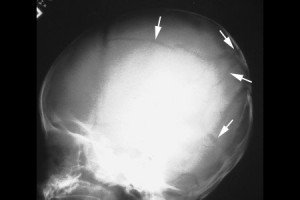
A simple linear skull fracture is a break in a cranial bone resembling a thin line, without splintering, depression, or distortion of bone. They are equally prevalent in NAI and in accidental injury. The picture below is of a linear right parietal skull fracture and comes from a Rumanian educational website, http://www.medandlife.ro/medandlife602.html.
This compares with a complex skull fracture which is variously defined as:
• a depressed fracture (where the skull is pushed in)
• two or more fractures of the skull
• fractures that cross the sutures (natural joining edges of skull bones) or those that are widening. (Source: Core-info leaflet)
The picture below of a complex skull fracture is from http://www.childabuseconsulting.com/child-abuse-fractures.html which houses other not-very-subtle images of non-accidental burns and bite marks as well.
Rib fractures in infants, particularly posterior ribs, with no history of major trauma are suspicious. The picture below is taken from http://www.learningradiology.com/notes/bonenotes/childabusepage.htm and shows multiple rib fractures with callous formation, the ones of the left 2nd and 6th posterior ribs being the easiest to identify:


