What constitutes sexualised behaviour in a 4 year old? This and the childhood asthma control test, this month, toddler fractures and the PCV vaccine. Do leave comments below.
Tag Archives: orthopaedics
2 months in one for Nov/Dec 2016
First part of information on gangs this month, plus HbA1c units compared, last bit on orthopaedic feet, a warning about phenytoin overdose and a couple of links to good relevant courses. Do leave comments below:
September 2016 uploaded
The value of PEWS this month, NICE on diabetes, a round up of former articles of use to the new batch of trainees and content from the paediatric orthopaedic team on funny shaped toes. Do leave comments below.
July 2016 PDF added
Malaria this month, sexual exploitation, sepsis, prolonged jaundice and DDH. Do leave comments below:
June 2016 published
Curly toes this month to herald the start of a new series on paediatric orthopaedics, sexual bullying, jaundice in the neonatal period and periorbital cellulitis. Do leave comments below…
November 2015 newsletter
November 2015: diagnosing asthma this month, a synopsis of vitamin D deficiency as we go into the winter, a helpful cartoon around mental well-being and hypermobility demystified. All comments gratefully received!
Potted background, assessment and management of vitamin D deficiency
Vitamin D deficiency in children with thanks to Dr Jini Haldar, paediatric registrar at Whipps Cross University Hospital.
Introduction
Vitamin D is an essential nutrient needed for healthy bones, and to control the amount of calcium in our blood. There is recent evidence that it may prevent many other diseases. There are many different recommendations for the prevention, detection and treatment of Vitamin D deficiency in the UK. The one outlined below is what we tend to do at Whipps Cross Hospital.
Prevention
The Department of Health and the Chief Medical Officers recommend a dose of 7-8.5 micrograms (approx. 300 units) for all children from six months to five years of age. This is the dose that the NHS ‘Healthy Start’ vitamin drops provide. The British Paediatric and Adolescent Bone Group’s recommendation is that exclusively breastfed infants receive Vitamin D supplements from soon after birth. Adverse effects of Vitamin D overdose are rare but care should be taken with multivitamin preparations as Vitamin A toxicity is a concern. Multivitamin preparations often contain a surprisingly low dose of Vitamin D.
Indications for measurement of vitamin D
1. Symptoms and signs of rickets/osteomalacia
- Progressive bowing deformity of legs
- Waddling gait
- Abnormal knock knee deformity (intermalleolar distance > 5 cm)
- Swelling of wrists and costochondral junctions (rachitic rosary)
- Prolonged bone pain (>3 months duration)
2. Symptoms and signs of muscle weakness
- Cardiomyopathy in an infant
- Delayed walking
- Difficulty climbing stairs
3. Abnormal bone profile or x-rays
- Low plasma calcium or phosphate
- Raised alkaline phosphatase
- Osteopenia or changes of rickets on x-ray
- Pathological fractures
4. Disorders impacting on vitamin D metabolism
- Chronic renal failure
- Chronic liver disease
- Malabsorption syndromes, for example, cystic fibrosis, Crohn’s disease, coeliac disease
- Older anticonvulsants, for example, phenobarbitone, phenytoin, carbamazepine
5. Children with bone disease in whom correcting vitamin D deficiency prior to specific treatment would be indicated:
- Osteogenesis imperfecta
- Idiopathic juvenile osteoporosis
- Osteoporosis secondary to glucocorticoids, inflammatory disorders, immobility
Symptoms and signs in children of vitamin D deficiency
1. Infants: Seizures, tetany and cardiomyopathy
2. Children: Aches and pains: myopathy causing delayed walking; rickets with bowed legs, knock knees, poor growth and muscle weakness
3. Adolescents: Aches and pains, muscle weakness, bone changes of rickets or osteomalacia
Risk factors for reduced vitamin D levels include:
- Dark/pigmented skin colour e.g. black, Asian populations
- Routine use of sun protection factor 15 and above as this blocks 99% of vitamin D synthesis
- Reduced skin exposure e.g. for cultural reasons (clothing)
- Latitude (In the UK, there is no radiation of appropriate wavelength between October and March)
- Chronic ill health with prolonged hospital admissions e.g. oncology patients
- Children and adolescents with disabilities which limit the time they spend outside
- Institutionalised individuals
- Photosensitive skin conditions
- Reduced vitamin D intake
- Maternal vitamin D deficiency
- Infants that are exclusively breast fed
- Dietary habits – low intake of foods containing vitamin D
- Abnormal vitamin D metabolism, abnormal gut function, malabsorption or short bowel syndrome
- Chronic liver or renal disease
Management depends on the patient’s characteristics:
A. No risk factors
No investigations, lifestyle advice* and consider prevention of risk factors
B. Risk Factors Only
1. Children under the age of 5 years: Lifestyle advice* and vitamin D supplementation.
Purchase OTC or via Healthy Start
Under 1 year: 200 units vitamin D once daily
1 – 4 years: 400 units vitamin D once daily
2. Children 5 years and over – offer lifestyle advice*
C. Risk Factors AND Symptoms, Signs
Lifestyle advice*
Investigations:
- Renal function, Calcium, Phosphate, Magnesium (infants), alkaline phosphatase,
- 25-OH Vitamin D levels, Urea and electrolytes, parathyroid hormone
Children can be managed in Primary Care as long as:
- No significant renal impairment
- Normal calcium (If <2.1 mmol/l in infants, refer as there is a risk of seizures)
If further assessment is required consider referral to specialist. **
Patient’s family is likely to have similar risk of Vitamin D deficiency – consider investigation ant treatment if necessary.
*Life style advice
1. Sunlight
Exposure of face, arms and legs for 5-10 mins (15-25 mins if dark pigmented skin) would provide good source of Vitamin D. In the UK April to September between 11am and 3pm will provide the best source of UVB. Application of sunscreen will reduce the Vitamin D synthesis by >95%. Advise to avoid sunscreen for the first 20-30 minutes of sunlight exposure. Persons wearing traditional black clothing can be advised to have sunlight exposure of face, arms and legs in the privacy of their garden.
2. Diet
Vitamin D can be obtained from dietary sources (salmon, mackerel, tuna, egg yolk), fortified foods (cow, soy or rice milk) and supplements. There are no plant sources that provide a significant amount of Vitamin D naturally.
**Criteria for referral
- Criteria for management in primary care not met
- Deficiency established with absence of known risk factors
- Atypical biochemistry (persistent hypophosphatemia, elevated creatinine)
- Failure to reduce alkaline phosphatase levels within 3 months
- Family history (parent, siblings) with severe rickets
- Infants under one month with calcium <2.1mmmol/l at diagnosis as risk of seizure. (Check vitamin D level of mothers in this group immediately and treat, particularly if breast feeding.)
- If compliance issues are anticipated or encountered during treatment.
- Satisfactory levels of vitamin D not achieved after initial treatment.
Vitamin D levels, effects on health and management of deficiency
| level | effects |
management |
| < 25 nmol/l (10micrograms/l) | Deficient. Associated with rickets, osteomalacia | Treat with high dose vitamin D
Lifestyle advice AND vitamin D (ideally cholecalciferol) • 0 – 6 months: 3,000 units daily • 6 months – 12 yrs: 6,000 units daily • 12 – 18 yrs: 10,000 units daily |
| vitamin D 25 – 50 nmol/l (10 – 20micrograms/l | Insufficient and associated with disease risk | Over the counter (OTC) Vitamin D supplementation (and maintenance therapy following treatment for deficiency) should be sufficient.
• Lifestyle advice and vitamin D supplementation < 6 months: 200 – 400 units daily (200 units may be inadequate for breastfed babies) Over 6 months – 18 years: 400 – 800 units daily |
| 50 – 75 nmol/l (20 – 30micrograms/l) | Adequate | Healthy Lifestyle advice |
| > 75 nmol/l (30 micrograms/l) | Optimal Healthy | None |
Course length is 8 – 12 weeks followed by maintenance therapy.
Checking of levels again
As Vitamin D has a relatively long half-life levels will take approximately 6 months to reach a steady state after a loading dose or on maintenance therapy. Check serum calcium levels at 3 months and 6 months, and 25 – OHD repeat at 6 months. Review the need for maintenance treatment. NB: the Barts Health management protocol uses lower treatment doses for a minimum of 3 months and then there is no need for repeat blood tests in the majority of cases of children satisfying the criteria for management in primary care.
Serum 25 OHD after 3 months treatment Action
| level | action | review |
| >80nmol/ml | Recommend OTC prophylaxis and lifestyle advice | as required |
| 50 – 80 nmol/mL | Continue with current treatment dose | reassess in 3 months |
| < 50 nmol/mL | Increase dose or, in case of non-adherence/concern refer to secondary care. |
It is essential to check the child has a sufficient dietary calcium intake and that a maintenance vitamin D dose follows the treatment dose and is continued long term.
Follow-up:
Some recommend a clinical review a month after treatment starts, asking to see all vitamin and drug bottles. A blood test can be repeated then, if it is not clear that sufficient vitamin has been taken.
Current advice for children who have had symptomatic Vitamin D deficiency is that they continue a maintenance prevention dose at least until they stop growing. Dosing regimens vary and clinical evidence is weak in this area. The RCPCH has called for research to be conducted. The RCPCH advice on vitamin D is at http://www.rcpch.ac.uk/system/files/protected/page/vitdguidancedraftspreads%20FINAL%20for%20website.pdf
JINI HALDAR
How to use the Beighton score
Hypermobility
– with thanks to Dr Joe Ward, paediatric registrar at Whipps Cross University Hospital.
 |
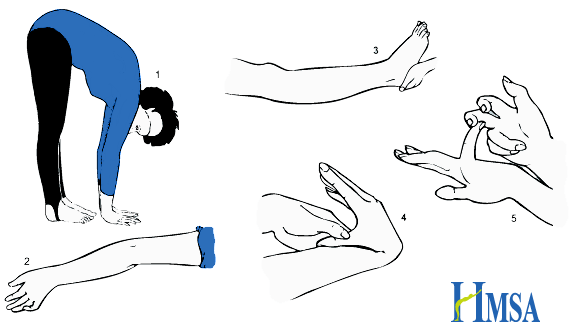
Picture from Hypermobility Syndromes Association
Hypermobility = synovial joints moving beyond normal range of movement.
Defined by the Beighton Score.: 1,2
- Ability to touch palms flat to floor with knees straight (one point)
- Elbow extension >10° (one point for each side)
- Knee extension >10° (one point for each side)
- Ability to touch thumb to forearm (one point for each side)
- Fifth finger metocarpalphalageal joint extension >90° (one point for each side)
Scores of 4 or above indicate Generalised Joint Hypermobility. May be asymptomatic, or associated with joint pain (exacerbated by exercise), dislocations and fatigue. Chronic pain often leads to muscle weakness. Other associations include dizziness and syncope and gastrointestinal problems such as chronic abdominal pain and constipation.
Physiotherapy and exercises to strengthen muscles around hypermobile joints provide the mainstay of treatment. Exercises to improve balance and coordination may also be helpful as proprioception may be impaired. Occupational therapy input may be beneficial.
The Brighton Criteria (NB: Brighton, not Beighton) is used in adults to diagnose Joint Hypermobility Syndrome. To make the diagnosis you need one of: two major criteria; one major and two minor criteria; four minor criteria; two minor criteria and one affected first degree relative. The presence of an underlying syndrome excludes the diagnosis. It is not yet validated in children.
Major Criteria:
- Beighton Score >3
- Arthralgia > 3 months in four or more joints
Minor Criteria:
- Beighton Score 1-3,
- Arthralgia > 3 months in one joint, backpain, or spondylosis / spondylolysis / spondylolisthesis
- Dislocation or subluxation in more than one joint, or in one joint repeatedly
- Three or more soft tissue lesions (e.g epicondylitis, tenosynovitis, bursitis)
- Marfanoid habitus
- Skin striae
- Ocular signs (e.g drooping eyelids, myopia, antimongoloid slant)
- Varicose veins, hernia, uterine or rectal prolapse
- Mitral valve prolapse
Connective tissue disorders associated with hypermobility should be excluded amongst children who meet the criteria for Joint Hypermobility Syndrome. These include but are not limited to:
Ehlers-Danlos Syndrome (http://www.ehlers-danlos.org/) – Heterogeneous group of disorders involving skin laxity, joint hypermobility, and vascular complications. Defined by the Villefranche Classification.
Marfan’s Syndrome (http://www.marfan.org/about/marfan) – Autosomal dominant connective tissue disorder. Typical features include characteristic facies, joint laxity, musculoskeletal problems (bone overgrowth and disproportionately long limbs), lens dislocation, and cardiovascular complications including aortic root dilatation.
Further information:
References:
1. Cattalini, M., Khubchandani, R. & Cimaz, R. When flexibility is not necessarily a virtue: a review of hypermobility syndromes and chronic or recurrent musculoskeletal pain in children. Pediatr. Rheumatol. Online J. 13, (2015).
2. Pacey, V., Tofts, L., Wesley, A., Collins, F. & Singh-Grewal, D. Joint hypermobility syndrome: a review for clinicians. J. Paediatr. Child Health 51, 373–380 (2015).
April and May!
I seem to have forgotten to put a blog post up when I published April’s newsletter which contains information on: tonsillectomy for parents, erythema infectiosum (which I think my son had this week), a safety alert about bath seats, tranexamic acid in paediatric trauma and how to make a nasal douche for rhinitis sufferers.
May is now also published and features dangerous dogs, knee pain, dental caries and continuations of both the dermatology and ENT features. Do leave comments below.
March 2014 newsletter
March brings urticaria, headaches, rugby injuries, Severs disease and bruising. Do leave comments below: