Category Archives: For Emergency Departments

Foreign body ingestion

The information for this topic is taken from a recent comprehensive review (August 2012) that appeared in www.UpToDate.com.  Bartshealth employees can access the full text via a link from the intranet.

Ingested foreign bodies (UptoDate.com article, August 2012)

Coins — Coins are by far the most common foreign body ingested by children. Approximately two-thirds of ingested coins are in the stomach by the time of x-ray but those that lodge in the oesophagus for 24 hours after ingestion may need to be removed endoscopically as only 20-30% of these will pass into the stomach on their own.  Coins that reach the stomach can be managed expectantly, and most will be passed within one to two weeks. A child who develops any signs or symptoms of obstruction, abdominal pain, vomiting, or fever, needs to come back to the ED urgently.

Button batteries — ingestions of “button” batteries are increasing and are associated with significant morbidity. Animal studies have demonstrated mucosal necrosis within one hour of ingestion and ulceration within two hours, with perforation as early as eight hours after ingestion.  It may be difficult to differentiate between a disk battery and a coin on a radiograph. This distinction is most important when the foreign body is in the oesophagus, since batteries require immediate removal whereas coins may not.

Magnets — also increasing. Many of the children with complications from multiple magnet ingestion had underlying developmental delay or autism. In one case, an older child inadvertently swallowed these magnets while using them to imitate a pierced tongue.  Two or more strong magnets, especially if ingested at different times, may attract across layers of bowel leading to pressure necrosis, fistula, volvulus, perforation, infection, or obstruction. Radiographs of the neck and abdomen should be performed, including a lateral view. X-rays cannot usually determine whether bowel wall is compressed between the magnets, although the finding of magnets that appear to be stacked but are slightly separated is suggestive. Management depends on the number, location and type of magnets, and on the timing of the ingestion.  Ingestion of a single magnet can generally be managed conservatively with serial radiographs while multiple magnets need removing.  Laxatives may help with faster bowel emptying if they are not in a place easily accessible with the endoscope.

References at www.uptodate.com.


September 2012 newsletter!

Take a look at September 2012’s edition of Paediatric Pearls!  Safeguarding issues surrounding head and spinal injuries, simple motor tics, chronic fatigue syndrome, the new CATS website and some pointers to gems you might have missed from the last 3 years.  Do leave comments.

child abuse and head injuries

This summarises the Core-info leaflet on head and spinal injuries in children. Full details are available at www.core-info.cardiff.ac.uk.

**PLEASE REFER ALL SUSPECTED INFLICTED HEAD AND SPINAL INJURIES TO PAEDIATRICS **

Inflicted head injuries

  • can arise from shaking and/or impact
  • occurs most commonly in the under 2’s
  • are the leading cause of death among children who have been abused
  • survivors may have significant long term disabilities
  • must be treated promptly to minimise long term consequences
  • victims often have been subject to previous physical abuse

Signs of inflicted head injury

  • may be obvious eg. loss of consciousness, fitting, paralysis, irritability
  • can be more subtle eg. poor feeding, excessive crying, increasing OFC
  • particular features include retinal haemorrhages, rib fractures, bruising to the head and/or neck and apnoeas
  • also look for other injuries including bites, fractures, oral injuries

If inflicted head injury is suspected

  • a CT head, skull X-ray and/or MRI brain should be performed
  • neuro-imaging findings include subdural haemorrhages +/- subarachnoid haemorrhages (extradural haemorrhages are
    more common in non-inflicted injuries)
  • needs thorough examination including ophthalmology and skeletal survey
  • co-existing spinal injuries should be considered
  • any child with an unexplained brain injury need a full investigation eg. for metabolic and haematological conditions, before a diagnosis of abuse can be made

The following diagram comes from http://www.primary-surgery.org:


These CT images are from http://www.hawaii.edu/medicine/pediatrics/pemxray/v5c07.html:


EXTRADURAL (or epidural) haematoma


SUBDURAL haemorrhages in a 4 month old

SUBARACHNOID haemorrhage in a 14 month old

Neuro-imaging for inflicted brain injury should be performed in

  • any infant with abusive injuries
  • any child with abusive injuries and signs and symptoms of brain injury

Inflicted spinal injuries

  • come in 2 categories : neck injuries, and chest or lower back injuries
  • neck injuries are most common under 4 months
  • neck injuries are often associated with brain injury and/or retinal haemorrhages
  • chest or lower back injuries are most common in older toddlers over 9 months
  • if a spinal fracture is seen on X-ray or a spinal cord injury is suspected, an MRI should be performed


August 2012 PDF digest

August’s PDF only has 4 text boxes but with lots of information crammed into them and extra on the blog.  A great looking PDF on poisoning in children from one of our registrars, an article on stammering from another working with a speech and language therapist and an update on BTS pneumonia guidelines just in time for the winter.  Also a feature on Cardiff’s core info safeguarding work on the evidence behind different types of fractures.  Do leave comments…

Fractures in child abuse

Metaphyseal fractures, also known as a bucket handle, chip or corner fracture, occur at the growing end of the bone and only in children. Recent fractures are very difficult to see on x-ray and they are often not associated with any clinical sign of soft tissue swelling or bruising.  They may become more obvious radiographically after 11 to 14 days. They are thought to happen when the baby has been pulled or swung violently and the relatively weaker growing point of the bone breaks.  They have been noted to occur accidentally following birth injuries, following serial casting of talipes or as a consequence of appropriate physiotherapy to newborn babies. (Source: Core-info leaflet)

The picture of a metaphyseal fracture of an infant’s wrist below comes from a 2000 paper on the orthopaedic aspects of child abuse by Kocher et al and published in the Journal of the American Academy of Orthopaedic Surgeons (http://www.jaaos.org/content/8/1/10.abstract):


A spiral fracture refers to the direction in which the bone is fractured.  It implies that there has been a twisting force to cause the fracture. Spiral fractures can also occur accidentally in the femur once the child is walking. (Source: Core-info leaflet)

The picture below of a spiral fracture of the femur in a 2 month old comes from http://www.hawaii.edu/medicine/pediatrics/pemxray/v6c02.html (same website has a wealth of other paediatric radiological images on it if you are interested):


A supracondylar fracture is one in the upper arm immediately above the elbow and is highly suggestive of accidental injury.  The picture below comes from http://www.kidsfractures.com/, a site put together by 2 American orthopaedic surgeons who say their aim was to make parents’ experience of having a child with a broken bone a little less traumatic but I think much of the language and many of the pictures are more suited to a medical audience.


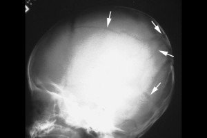
A simple linear skull fracture is a break in a cranial bone resembling a thin line, without splintering, depression, or distortion of bone.  They are equally prevalent in NAI and in accidental injury.  The picture below is of a linear right parietal skull fracture and comes from a Rumanian educational website, http://www.medandlife.ro/medandlife602.html.


This compares with a complex skull fracture which is variously defined as:

• a depressed fracture (where the skull is pushed in)

• two or more fractures of the skull

• fractures that cross the sutures (natural joining edges of skull bones) or those that are widening. (Source: Core-info leaflet)

The picture below of a complex skull fracture is from http://www.childabuseconsulting.com/child-abuse-fractures.html which houses other not-very-subtle images of non-accidental burns and bite marks as well.


Rib fractures in infants, particularly posterior ribs, with no history of major trauma are suspicious.  The picture below is taken from http://www.learningradiology.com/notes/bonenotes/childabusepage.htm and shows multiple rib fractures with callous formation, the ones of the left 2nd and 6th posterior ribs being the easiest to identify:


BTS 2011 guideline on community acquired pneumonia in children

In October 2011 the British Thoracic Society updated its guidelines on community acquired pneumonia in children.  Dr Michael Eyres looked at it in more detail for Paediatric Pearls.  He was also part of our local audit team contributing to the national audit.  The results showed that we, despite insisting on as few investigations as possible, are still doing too many chest x-rays, blood cultures and CRP measurements.  Think – will it change management?

Here are the basics:

When to consider pneumonia

Persistent fever > 38.5°C     +     chest recessions    + tachypnoea

Investigations

• CXR should not be considered routine and is not required in children who do not need admission.

• Acute phase reactants including CRP are not useful in distinguishing viral from bacterial infection and should not
be tested routinely. Blood cultures also do not need to be routinely taken.

• Daily U&Es are required in children receiving IV fluids.


Severity assessment

• Children with oxygen saturations <92% need hospital referral.

• Auscultation findings of absent breath sounds with dullness to percussion need hospital referral.

• Children should be reassessed if symptoms persist.


General management

• Give parents information on managing fever, preventing dehydration and identifying deterioration.

• Children with oxygen saturations <92% need oxygen.

• NG tubes should be avoided in severe respiratory compromise and in infants.

• Chest physio is not beneficial and should not be performed in pneumonia.


Antibiotics

• All children with a clear clinical diagnosis of pneumonia should receive antibiotics as bacterial and viral
infections cannot be reliably distinguished. However most children younger than 2 years presenting with mild symptoms of respiratory distress (this would
include the bronchiolitics) do not usually require antibiotics.

• Amoxicillin is the oral first-line for all children as it is effective, well tolerated and cheap.

• Macrolides if no response to first-line / suspected mycoplasma or chlamydia / very severe disease.

• Augmentin if pneumonia associated with influenza.

• Oral agents are effective even in severe pneumonia; IV is needed only if unable to tolerate oral or there are
signs of septicaemia, empyema or abscess.


Follow-up

• Children with severe pneumonia or complications should be followed up after discharge until they have recovered completely and
CXR is near normal. Follow-up CXR is not otherwise required, but may be considered in round pneumonia, collapse or if symptoms persist.


June 2012 PDF

June’s PDF digest is ready for consumption.  Both APLS and NICE have lost paraldehyde from their status epilepticus algorithms, a link to Working Together and an article on sticky eyes v. conjunctivitis.  Blood pressure centile charts and a plug for the London Deanery’s communication skills courses.  Do leave comments below.

May 2012 ready a week early!

Click here for May’s PDF digest of Paediatric Pearls.  Perinatal infant-maternal mental health this month from both SIGN and our local services, diarrhoea and vomiting fluid calculation, pertussis, bruise assessment in NAI and food allergy.  Also a couple of links to useful parenting support sites.  Do leave comments!

The Perinatal Parent Infant Mental Health Service (PPIMHS)

The PPIMHS teams are made up of perinatal psychiatrists, community mental health practitioners and psychotherapists/psychologists and they accept referrals from Health Visitors, GPs, midwives, Children’s Centres workers or other health professionals and self-referrals.  Click here for their referral form.  They may signpost elsewhere after the initial consultation if appropriate or they will offer the parent/carer and infant/child 9-12 sessions to work on the parent-infant relationship and/or psychiatric support as required.

Groups particularly at risk of having problems with bonding include families with ex-premature babies who have spent a significant amount of time on the Special Care Baby Unit, those where the baby has feeding issues or is difficult to soothe, those where breastfeeding failed to establish and those where there was a traumatic birth or difficult conception and/or pregnancy.  Many of the parents on their case load have a personal history of disturbed attachments and are keen not to let history repeat itself.  A recent audit showed that 41% of their mothers had some sort of mental health diagnosis which means that 59% did not.  Click here for an information leaflet about their service that you might like to give to your patients.

Mums with postnatal depression or post-partum psychosis should be referred directly to PPIMHS.  Parents struggling with a crying baby or fussy toddler but with no bonding issues should be referred to their health visitor.  The PPIMHS team is a tier 3 (specialised) service concentrating primarily on the parent-infant relationship and perinatal mental health.

Symptoms in the baby that might suggest a bonding problem:

extreme clingy behaviours, fussy, difficult to soothe, abnormal self-soothing behaviours (eg. head-banging, hair-pulling, scratching), excessive sleep problems, extreme feeding problems, lack of verbal and non-verbal communication, stiff or floppy posture, extreme fearfulness or watchfulness, lack of interest in the world, no comfort sought from parents, avoids eye contact with parents, smiles very little.

Symptoms in the parent:

high anxiety and panic about the baby, excessive A and E or GP presentations, feeling frightened of harming the baby, lack of separation between parent and baby, baby never put down, excessive sterilising of bottles and toys, detached feelings about the baby, no pride in their development, anger about baby as if baby intends to upset the parent, feelings of failure as a parent, inability to cope.

There is some evidence around this issue and around maternal stress during pregnancy and the effect of high maternal cortisol levels on the foetus’ developing brain.  I have asked the Waltham Forest PPIMHS psychologists to write a bit about that and correct anything I have written about their service!