Category Archives: For Emergency Departments

Oral rehydration guideline

Most children who are dehydrated presenting to UK emergency departments can be rehydrated orally. 

  • Give 50ml/kg ORS solution over 4hrs, plus ORS solution for maintenance, often and in small amounts (even by syringe or spoon)
  • Continue breast feeding
  • Consider supplementing with usual fluids (but not fruit juices or carbonated drinks) if a child without red flag symptoms or signs (see http://www.nice.org.uk/CG84) refuses to take sufficient ORS solution.  Don’t give solids.
  • Consider giving ORS solution via ng tube if child is unable to take it or continues to vomit (esp. with red flag symptoms/signs)
  • Monitor carefully

This is a worked example for a 3 year old child weighing 14kgs who has been assessed as about 5% dehydrated.

Maintenance = 100mls/kg for first 10kgs and 50mls/kg for next 10 kgs = 1000mls + 200mls = 1200mls over 24 hours

Replacement = 5 x 14 x 10 = 700mls over the first 4 hours (extra to maintenance needs)

Therefore the child needs 225mls per hour for the first 4 hours (1200/24 + 700/4), followed by 50mls (1200/24) per hour.

The 225 mls is best given as 18 mls every 5 minutes or 56mls every 15 minutes if vomiting seems to have stopped or if using nasogastric tube.

They should have 5mls/kg = 70mls extra diarolyte (ORS) with each diarrhoeal stool or vomit.

Give parents written information to go home with so they understand that diarrhoea may continue for a few days but this does not matter as long as they are able to get enough fluid in the top end.  The NICE guideline parent information is at http://guidance.nice.org.uk/CG84/PublicInfo/pdf/English.

Whooping cough outbreak 2012

More background to pertussis with thanks to Dr Rupa Vora

  • whooping cough is caused by Bordetella pertussis, a gram negative pleomorphic bacillus. It is spread by aerosol transmission and the bacteria cause damage by attaching to the respiratory cilia
  • it occurs in clusters every 2-5 years during the summer months. We currently have an outbreak with the HPA provisionally reporting 665 cases in the first quarter of 2012 (cf. 1040 cases in 2011, 421 in 2010)
  • cases have dropped dramatically since pertussis vaccinations have been introduced. Acellular pertussis vaccination is given at 2 and 3 months, followed by a pre-school booster.  However, protection wanes quickly and has virtually disappeared by 12 years old
  • incubation period is 3-12 days and children are most infectious in the first 2-3 weeks. They are most likely to present in the second phase of illness at 3-4 weeks
  • can present with coryza (1st stage which lasts a couple of weeks), paroxysms of cough, difficulty feeding and pneumonia. Younger infants (<6months) may not present with the characteristic ‘whoop’. Older children and adults often present with a persistent cough
  • complications include chronic cough (“100 day cough”), hypoglycaemia, seizures, encephalopathy and intracranial haemorrhage
  • any infant is vulnerable and up to 50% may need hospitalisation.  Especially vulnerable are ex-prems and those with underlying cardiology, respiratory or neurological problems.  
  • In England and Wales, whooping cough is statutorily notifiable.  The diagnosis is usually made on clinical grounds without the requirement for laboratory confirmation
  • The UK Health Protection Agency advises a 7 day course of erythromycin or clarithromycin (or azithromycin for 3-5 days if under 4 weeks) to reduce spread.  A pernasal swab to confirm or refute B. pertussis as the causative organism can be carried out.  If the cough has been present for more than two weeks and the child is in the community, serum serology can be sent to Colindale.  See table below:


Appropriate laboratory tests for a sporadic case of pertussis reported to HPA on clinical suspicion (with thanks to Dr Maria O’Callaghan): 

Age Clinical symptoms
≤ 2 weeks cough > 2 weeks cough
≤ 1 yr

Hospitalised

NPA/PNS for PCR (RSIL)

PNS for culture (local laboratory)

NPA/PNS for PCR (RSIL)

PNS for culture (local laboratory)

Serum for serology (RSIL)

≤ 1 yr

community

PNS for culture (local laboratory) Serum for serology (RSIL)
> 1 yr to 6 yr
6 to 15 yr Serum for serology (RSIL)
> 15 yr

 NPA – nasopharyngeal aspirate; PNS – pernasal swab;

RSIL – Respiratory and Systemic Infections Laboratory, Colindale

Useful websites:

HPA: www.hpa.org.uk/Topics/InfectiousDiseases/InfectionsAZ/WhoopingCough/

NHS Choices: www.nhs.uk/Conditions/Whooping-cough/Pages/Introduction.aspx

Joint March and April edition of PP published!

With apologies for missing March – time management issues….

Functional abdominal pain this month with a link to a handy patient information leaflet, pubertal and growth issues, neonatal spots and some not-for-the-faint-hearted youtube videos on intraosseous line insertion for the ED practitioners amongst you.  Do leave comments!

Paediatric Pearls for February 2012

Click here for this month’s PDF digest!  It ‘s quite hard providing a balance of information for GPs and ED juniors now that I am only doing the one newsletter.  I think we’ve succeeded this month with neurodevelopmental milestones in Down’s syndrome and essential tremor aimed mainly at GPs and pulled elbow, anaphylaxis and the FEAST study aimed more towards the emergency medicine practitioners.  Many thanks to my colleagues who have contributed this month.  The FEAST video makes fascinating and inspiring watching for any health professional, regardless of specialty.  Do leave comments, questions, suggestions!

NICE on anaphylaxis

With thanks to my colleague, Dr Su Li, for summarising this 2011 NICE guideline for Paediatric Pearls.

Anaphylaxis: assessment to confirm an anaphylactic episode and the decision to refer after emergency treatment for a suspected anaphylactic episode

December 2011

www.nice.org.uk/cg134

Anaphylaxis is a severe, life-threatening, generalised hypersensitivity reaction involving

  • the airway (pharyngeal or laryngeal oedema) and/or
  • breathing (bronchospasm, tachypnoea) and/or
  • circulation (hypotension, tachycardia).


There can often be skin and mucosal changes. Patients presenting with these signs and symptoms should be diagnosed as having ‘suspected anaphylaxis’.

Anaphylaxis may be an allergic response that is

  • immunologically IgE mediated (foods, venoms, drugs, latex) or
  • non-immunologically mediated or
  • idiopathic (significant clinical effects with no obvious cause).


This guideline does not make any drug recommendations. These can be found at http://www.resus.org.uk/pages/reaction.pdf.

Patient Centred Care

  • Treatment and care should take into account patient’s needs and preferences
  • Patients should have the opportunity to make informed decisions about their care and treatment, in partnership with health care professionals
  • Good communication between healthcare professionals and patients is essential
  • Families and carers should be given the information and support they need
  • Care of young people in transition between paediatric and adult services should be planned and managed according to the best practice guidance described in ‘Transition: getting it right for young people


Recommendations

  • Document acute clinical features of the suspected anaphylaxis
  • Record the time of onset
  • Record the circumstances immediately before the onset of symptoms to help identify possible triggers


  • Consider taking blood samples for mast cell tryptase if reaction is thought to be immunologically mediated or idiopathic
    • First sample as soon after emergency treatment given
    • Second sample 1-2 hours (no more than 4 hours) from onset of symptoms
    • A further sample may be required at follow up with the allergy specialist to measure baseline mast cell tryptase


  • Children who have had emergency treatment should be admitted to hospital under the care of the paediatric team.  The resus council suggests observing the child for a pragmatic (no evidence yet) 6 hours because of the risk of a biphasic reaction.
  • Offer the child/parents a referral to an allergy specialist (see www.bsaci.org for registered allergy clinics)
  • Offer the child/parents an adrenaline injector in the interim period whilst waiting for a specialist appointment


  • Before discharge, offer the child/parents
    • Information about anaphylaxis (signs, symptoms, risk of recurrence of symptoms (biphasic reaction)).  Parent information leaflet here.
    • Information about what to do if a reaction occurs (use adrenaline injector, call emergency services)
    • Demonstration on how to use an adrenaline injector see http://www.youtube.com/watch?v=pgvnt8YA7r8 for a clear American description of how to use it.
    • Advice about how to avoid potential triggers
    • Information about the need for referral and the referral process to an allergy specialist
    • Information about patient support groups


Research Recommendations

  • Mast cell tryptase is not always elevated in children, particularly if food is thought to be the allergen or if respiratory compromise is the main clinical feature. It is recommended that further studies be carried out to identify other potential chemical inflammatory mediators.
  • There is limited evidence on biphasic reactions. Follow up studies are recommended.
  • There are no studies on length of observation period following emergency treatment for suspected anaphylaxis
  • There is limited data on the annual incidence or anaphylactic reactions and their associated outcomes.
  • The Guideline Development Group feel that referral to specialist services and/or the provision of adrenaline injectors are likely to benefit patients who have experienced a suspected anaphylaxis as a result of decreased anxiety and ongoing support. This benefit is yet to be quantified.

Pulled elbow

Dr Furqan Ahmed is an Emergency Medicine middle grade doctor seconded to paediatrics for a few months as part of his training.  I hope he is learning from us, we are certainly picking up things from him.  He has put together the following guide to “pulled elbow” or “radial head subluxation” for Paediatric Pearls.

Pulled elbow, Nursemaid’s elbow, is a dislocation of the elbow joint caused by a sudden pull on the extended, pronated arm. The technical term for the injury is radial head subluxation.

Pathophysiology:

The etiology is slippage of the head of the radius under the annular ligament. The distal attachment of the annular ligament covering the radial head is weaker in children than in adults, allowing it to be more easily torn.

As children age, the annular ligament strengthens, making the condition less common. The oval shape of the proximal radius in cross-section contributes to this condition by offering a more acute angle posteriorly and laterally, with less resistance to slippage of the ligament when axial traction is applied to the extended and pronated forearm.

Causes, incidence, and risk factors

Radial head subluxation is a common pediatric presentation generally occurring between the ages of 1 and 4 years, although it can happen anytime between 6 months of age and 7 years. After age 3, children’s joints and ligaments gradually grow stronger, making radial head subluxation less likely to occur.

The injury occurs when a child is pulled up too hard by the hand or wrist. It is often seen after someone lifts a child up by one arm (eg. when trying to lift the child over a curb or high step).

Other ways this injury may happen include:

  • Breaking a fall with the arm
  • Rolling over in an unusual way
  • Swinging a young child from the arms while playing


Signs and symptoms

When the injury occurs, the child usually begins crying right away and refuses to use the arm because of elbow pain.

  • The child may hold the arm slightly bent (flexed at 15-20 degrees) at the elbow and pressed up against the abdominal area (pronated).
  • The child will move the shoulder, but not the elbow. Some children stop crying as the first pain goes away, but continue to refuse to move the elbow.
  • Tenderness at the head of the radius may be present.
  • Erythema, warmth, oedema, or signs of trauma are absent.
  • Distal circulation, sensation, and motor activity are normal

Treatment

Inform child and caregiver that the reduction may be uncomfortable, but the discomfort will end quickly after reduction. Parents should not attempt these manoeuvres at home unless advised by a physician.

To resolve the problem, the affected arm must be held with one hand/finger on the radial head and the other grasping the hand making sure the elbow is in 90° of flexion. While applying compression between these two hands, the forearm of the patient is gently supinated and the arm flexed. The manipulator will usually feel a “click” if the manoeuvre is done properly, the child will feel momentary pain, and usually within 5 minutes, the forearm will be functioning well and painlessly.  NB: although a ‘click’ signifies reduction, absence of a ‘click’ is noted in some successful reductions.

Differential diagnoses:

  • Fracture, Elbow
  • Fracture, Wrist
  • Hand Injury, Soft Tissue


Indication for xray:

Child not using arm 30 minutes after a reduction.  External signs of trauma such as swelling, abrasions, or ecchymoses.

Consultations

If radiographic findings demonstrate no fracture, repeat attempts at reduction are unsuccessful, and the child does not regain normal function after 30-40 minutes, the safest management is to support the arm in a sling (or splint and sling) and have the child reevaluated in 1-2 days time.

Prognosis

The prognosis is excellent. Parents can be reassured that no permanent injury results from this condition.

For those who have had one occurrence, the chance of recurrence is approximately 20-25%.  Those 24 months and younger may have the greatest risk of recurrence.


References

  1. ^ Krul M, van der Wouden JC, van Suijlekom-Smit LW, Koes BW (2012). “Manipulative interventions for reducing pulled elbow in young children”. Cochrane Database Syst Rev (1): CD007759. doi:10.1002/14651858.CD007759.pub3. PMID 22258973
  2. ^ Toupin P, Osmond MH, Correll R, Plint A (September 2007). “Radial head subluxation: how long do children wait in the emergency department before reduction?”. CJEM 9 (5): 333–7. PMID 17935648. http://www.cjem-online.ca/v9/n5/p333
  3. ^ Kaplan, RE; Lillis, KA (2002 Jul). “Recurrent nursemaid’s elbow (annular ligament displacement) treatment via telephone.”. Pediatrics 110 (1 Pt 1): 171–4. PMID 12093966
  4. ^ Macias CG, Bothner J, Wiebe R (July 1998). “A comparison of supination/flexion to hyperpronation in the reduction of radial head subluxations”. Pediatrics 102 (1): e10. PMID 9651462. http://pediatrics.aappublications.org/cgi/pmidlookup?view=long&pmid=9651462.

January 2012 PDF ready

Do you know your valgus from your varus?  Or your myclonic epilepsy from your sleep myoclonus?  A link this month to new asthma patient information leaflets and some reminders of NICE’s “Do not do recommendations” in feverish children.  Also the BSACI egg allergy guideline.  Do leave comments on any of these topics below.

Antipyretics – single or dual therapy?

Managing Fever in Children with thanks to Dr Ranjev Kainth

Fever, both in primary and secondary care is a frequent presentation. Often, it is a sign of an illness and in the first instance, it is important to establish the most likely underlying causative factor. Once this has been determined, focus often turns to the management of the fever.  Two recent articles in Archives highlight the varying practice amongst clinicians both in primary and secondary care.

In accordance with NICE guidelines1, the authors2-3 suggest anti-pyretics should not be used for the sole purpose of controlling fever.  Agents such as paracetamol and ibuprofen are often administered to promote comfort in the child when there is  fever.  In such situations, children may be prescribed single or dual therapy.

In a systematic review, E.Purssell3 examines the evidence for combined anti-pyretic therapy with either paracetamol or ibuprofen alone. He concluded that ‘only marginal benefit was shown for the combined treatment compared with each drug individually which, taken alongside the risk of overdose and further increasing the fear of fever, suggests there is little to recommend this practice’.

With the real risk of parents being unable to accurately measure medication4, it is important clear guidance is given on when and what type of drug therapy is appropriate in clinical situations.

References:

  1. NICE Guideline: Feverish Illness in Children:  Quick reference guideline http://guidance.nice.org.uk/CG47/QuickRefGuide/pdf/English
  2. McLyntyre, J. Management of fever in Children. Arch Dis Child Dec 2011 V 96;12 P.1173
  3. Purssell, E. Systematic review of studies comparing combined treatment with paracetamol and ibuprofen, with either drug alone. Arch Dis Child Dec 2011 V 96;12 P.1175
  4. Beckett, V.L. et al. Accurately administering oral medication to children isn’t child’s play. Arch Dis Child 2011;96:A7 doi:10.1136/adc.2011.212563.14  

December 2011. Happy Christmas!

December 2011 has snippets of information on torticollis (backed up with lots more information on the website), unconscious children, alkaline phosphatase and a link to the Map of Medicine’s recent algorithm for cough in children.  Also some pointers for your safeguarding training needs.  Download it here.

Torticollis

Torticollis / Wry Neck / Sternomastoid tumour of infancy with thanks to Dr Katie Knight

(From Latin tortus = twisted + collum = neck)

Torticollis can be congenital or acquired, but this article will focus mostly on the congenital form, affecting 0.3% of infants and usually presenting in the first 6 months of life [1]. It is the third most common reason for referral to orthopaedics in this age group. The overwhelming majority of cases seen are due to a benign muscular problem, but some more sinister diagnoses can also present in a similar way, so it is crucial to be aware of these.

What causes torticollis?

Muscular damage:

Most cases of congenital torticollis are the result of damage to the sternocleidomastoid muscle (SCM) at birth (for example in instrumental delivery) or in the uterus (restricted movement or abnormal positioning causing muscle damage).

Damage to the SCM causes it to shorten or contract as fibrosis affects the area. Fibrotic change in the damaged muscle is felt as a hard lump – the ‘pseudotumour’ of torticollis, as it is sometimes called.

This shortening of the muscle in turn makes it difficult for the infant to turn their head, resulting in neck stiffness and a fixed head position, with very limited neck movement.

Risk of muscular torticollis is increased in intrauterine constraint (eg breech presentation or oligohydramnios [2]), and it is also associated with other minor positional deformities. 10% of babies with torticollis have hip dysplasia. [3] One study looking at 1001 babies found that 10% had one or more postural deformities (in decreasing order of frequency: plagiocephaly or torticollis; congenital scoliosis or pelvic obliquity; adduction contracture of a hip and/or malpositions of the knees or feet [4]. This study found that all these deformities were more likely to be observed in:

  • babies with a greater birth length
  • breech presentation
  • oligohydramnios
  • babies delivered instrumentally
  • Male infants were also found to be 1.9 x more likely to have positional deformities including torticollis.

With these presenting symptoms described above and nothing else of note, torticollis is clearly the first diagnosis that springs to mind. HOWEVER – to play the devil’s advocate – a baby who presents with ‘a lump in the neck’ and ‘abnormal neurology’ certainly demands a careful history and examination.

Uncommon causes of torticollis

Congenital vertebral abnormalities:

The SCM is supplied by the accessory nerve (CN XI), which exits the skull through the jugular foramen. Anything affecting the structure of the upper cervical spine or skull base could compress the nerve root of CN XI and cause torticollis.

Congenital vertebral abnormalities often come along with other congenital abnormalities, as part of a syndrome (two examples are briefly described below, for interest). For this reason a child presenting with torticollis who is known to have other congenital abnormalities should be carefully examined with the possibility of an unusual syndrome kept in mind. [5]

MURCS syndrome (Müllerian duct/renal aplasia/cervicothoracic somite dysplasia) is a rare condition affecting 1 in 5000 female infants that has been associated with congenital torticollis in some cases due to aplasia of the posterior vertebral arch [6]

Klippel-Feil syndrome – cervical spine fusion is seen along with a host of other symptoms [7]

Posterior fossa tumours, tumours of the cervical spine, atlas and axis – these are very rare and should be part of the differential in older children who present with acquired torticollis. [8, 9]  Posterior fossa tumours, when they present with torticollis, usually have accompanying symptoms of intracranial pathology (headache, nausea, vomiting, eye signs) [10]

‘Mimics’ of torticollis

‘Ocular torticollis’ occurs when there is 4th cranial nerve palsy. The superior oblique muscle, supplied by CN IV, causes the eye to look inwards and downwards. Paralysis of the muscle means the eye cannot adduct or internally rotate, and this causes torsional diplopia, which the child ‘corrects’ by tilting the head position. Adopting this position over a long period of time eventually causes contracture of the SCM. [11] This condition can be ruled out by using the cover test (watch a 7 minute long Youtube video with a rather disconcerting picture of a huge eye in the background here).  When the affected eye is covered, the child should spontaneously correct their head position (in the early stages, before muscle contracture has occurred).

Examination

Appearance (see image): The head is tilted to one side (to the side of the affected muscle), and the chin is turned to the other side. There is stiffness, from the lack of movement, so there may be pain when the neck position is passively corrected.

A lump may be felt in the distal SCM.
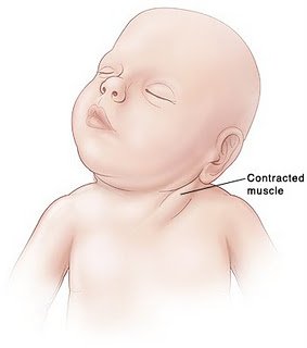
Lump felt in distal SCM


 Investigation

The key is to differentiate between muscular torticollis (ie common, benign, easily correctible) and non-muscular torticollis (ie possibly secondary to neurological, ocular or vertebral pathology, and needing further investigation.

If there is a lump palpable in the SCM, it needs to be differentiated from other causes of a lump in the neck. Ultrasound is the best first line investigation – it detects fibrosis of the muscle (diagnosing torticollis) but would also pick up abnormal lymph nodes or masses.

Fine needle aspiration would be the next step if there was any uncertainty of the diagnosis, but this is rarely needed.

 Treatment

 Once muscular torticollis is confirmed:

 Physiotherapy is the mainstay of treatment. Even when there is severe fibrosis of the SCM, physio is effective in 98% [12]

  •  Neck stretches, performed regularly, moving the neck in the opposite direction to the affected muscle (tilt head sideways towards non-affected side, rotate towards affected side). Physio referral is indicated so parents can be taught the correct way to perform the stretches.
  • Let the baby spend more time lying on its tummy, to strengthen neck muscles
  • Use baby chair or Fraser chair to minimise the time the baby spends lying flat
  • Encourage head turning to affected side by using toys, distraction, feeding from that side
  • Physiotherapist may advise use of a neck brace in certain cases.

 (The above advice adapted from ‘Physio Questions’ [13], a blog by an Australian physio – torticollis featured as a blog entry in August 2010)


Surgical treatment is very rarely needed – only in instances where conservative management has failed after 6 months of treatment. When surgery is performed, the operation is a bipolar release of the SCM, and this has been found to be highly successful, even in patients older than 5 years [14] and into adulthood [15]

Alternatives to surgery?

A recent successful non-surgical development in treating cases resistant to physio is using botox injection. [16] The evidence for chiropractic treatment is weak, isolated successful cases have been described, [17] but there has been no randomised controlled trial. There are also reports of infants with torticollis caused by neurogenic tumours being treated (unsuccessfully) by a chiropractor before the correct diagnosis was made, [18] so it is imperative that parents have consulted a doctor before they choose to seek chiropractic help.

 References:

  1. http://www.ncbi.nlm.nih.gov/pubmed/3566514?tool=bestpractice.bmj.com
  2. http://www.ncbi.nlm.nih.gov/pubmed/21376202
  3. http://www.ncbi.nlm.nih.gov/pubmed/7484683
  4. http://www.ncbi.nlm.nih.gov/pubmed/18795328
  5. http://web.jbjs.org.uk/cgi/reprint/71-B/3/404
  6. http://www.ncbi.nlm.nih.gov/pubmed/21553338
  7. http://emedicine.medscape.com/article/1264848-overview
  8. http://www.ncbi.nlm.nih.gov/pubmed/22095422
  9. http://www.ncbi.nlm.nih.gov/pubmed/20638308
  10. http://www.ncbi.nlm.nih.gov/pubmed/8784707
  11. http://www.ncbi.nlm.nih.gov/pubmed/868283
  12. http://www.ncbi.nlm.nih.gov/pubmed/21843719
  13. http://physioquestions.blogspot.com/2010/08/are-you-worried-about-your-childs.html
  14. http://www.ncbi.nlm.nih.gov/pubmed/22045346
  15. http://www.ncbi.nlm.nih.gov/pubmed/19036153
  16. http://www.ncbi.nlm.nih.gov/pubmed/16470158
  17. http://www.ncbi.nlm.nih.gov/pubmed/8263436
  18. http://www.ncbi.nlm.nih.gov/pmc/articles/PMC2484567/?tool=pmcentrez