What constitutes sexualised behaviour in a 4 year old? This and the childhood asthma control test, this month, toddler fractures and the PCV vaccine. Do leave comments below.
Tag Archives: fractures
Potted background, assessment and management of vitamin D deficiency
Vitamin D deficiency in children with thanks to Dr Jini Haldar, paediatric registrar at Whipps Cross University Hospital.
Introduction
Vitamin D is an essential nutrient needed for healthy bones, and to control the amount of calcium in our blood. There is recent evidence that it may prevent many other diseases. There are many different recommendations for the prevention, detection and treatment of Vitamin D deficiency in the UK. The one outlined below is what we tend to do at Whipps Cross Hospital.
Prevention
The Department of Health and the Chief Medical Officers recommend a dose of 7-8.5 micrograms (approx. 300 units) for all children from six months to five years of age. This is the dose that the NHS ‘Healthy Start’ vitamin drops provide. The British Paediatric and Adolescent Bone Group’s recommendation is that exclusively breastfed infants receive Vitamin D supplements from soon after birth. Adverse effects of Vitamin D overdose are rare but care should be taken with multivitamin preparations as Vitamin A toxicity is a concern. Multivitamin preparations often contain a surprisingly low dose of Vitamin D.
Indications for measurement of vitamin D
1. Symptoms and signs of rickets/osteomalacia
- Progressive bowing deformity of legs
- Waddling gait
- Abnormal knock knee deformity (intermalleolar distance > 5 cm)
- Swelling of wrists and costochondral junctions (rachitic rosary)
- Prolonged bone pain (>3 months duration)
2. Symptoms and signs of muscle weakness
- Cardiomyopathy in an infant
- Delayed walking
- Difficulty climbing stairs
3. Abnormal bone profile or x-rays
- Low plasma calcium or phosphate
- Raised alkaline phosphatase
- Osteopenia or changes of rickets on x-ray
- Pathological fractures
4. Disorders impacting on vitamin D metabolism
- Chronic renal failure
- Chronic liver disease
- Malabsorption syndromes, for example, cystic fibrosis, Crohn’s disease, coeliac disease
- Older anticonvulsants, for example, phenobarbitone, phenytoin, carbamazepine
5. Children with bone disease in whom correcting vitamin D deficiency prior to specific treatment would be indicated:
- Osteogenesis imperfecta
- Idiopathic juvenile osteoporosis
- Osteoporosis secondary to glucocorticoids, inflammatory disorders, immobility
Symptoms and signs in children of vitamin D deficiency
1. Infants: Seizures, tetany and cardiomyopathy
2. Children: Aches and pains: myopathy causing delayed walking; rickets with bowed legs, knock knees, poor growth and muscle weakness
3. Adolescents: Aches and pains, muscle weakness, bone changes of rickets or osteomalacia
Risk factors for reduced vitamin D levels include:
- Dark/pigmented skin colour e.g. black, Asian populations
- Routine use of sun protection factor 15 and above as this blocks 99% of vitamin D synthesis
- Reduced skin exposure e.g. for cultural reasons (clothing)
- Latitude (In the UK, there is no radiation of appropriate wavelength between October and March)
- Chronic ill health with prolonged hospital admissions e.g. oncology patients
- Children and adolescents with disabilities which limit the time they spend outside
- Institutionalised individuals
- Photosensitive skin conditions
- Reduced vitamin D intake
- Maternal vitamin D deficiency
- Infants that are exclusively breast fed
- Dietary habits – low intake of foods containing vitamin D
- Abnormal vitamin D metabolism, abnormal gut function, malabsorption or short bowel syndrome
- Chronic liver or renal disease
Management depends on the patient’s characteristics:
A. No risk factors
No investigations, lifestyle advice* and consider prevention of risk factors
B. Risk Factors Only
1. Children under the age of 5 years: Lifestyle advice* and vitamin D supplementation.
Purchase OTC or via Healthy Start
Under 1 year: 200 units vitamin D once daily
1 – 4 years: 400 units vitamin D once daily
2. Children 5 years and over – offer lifestyle advice*
C. Risk Factors AND Symptoms, Signs
Lifestyle advice*
Investigations:
- Renal function, Calcium, Phosphate, Magnesium (infants), alkaline phosphatase,
- 25-OH Vitamin D levels, Urea and electrolytes, parathyroid hormone
Children can be managed in Primary Care as long as:
- No significant renal impairment
- Normal calcium (If <2.1 mmol/l in infants, refer as there is a risk of seizures)
If further assessment is required consider referral to specialist. **
Patient’s family is likely to have similar risk of Vitamin D deficiency – consider investigation ant treatment if necessary.
*Life style advice
1. Sunlight
Exposure of face, arms and legs for 5-10 mins (15-25 mins if dark pigmented skin) would provide good source of Vitamin D. In the UK April to September between 11am and 3pm will provide the best source of UVB. Application of sunscreen will reduce the Vitamin D synthesis by >95%. Advise to avoid sunscreen for the first 20-30 minutes of sunlight exposure. Persons wearing traditional black clothing can be advised to have sunlight exposure of face, arms and legs in the privacy of their garden.
2. Diet
Vitamin D can be obtained from dietary sources (salmon, mackerel, tuna, egg yolk), fortified foods (cow, soy or rice milk) and supplements. There are no plant sources that provide a significant amount of Vitamin D naturally.
**Criteria for referral
- Criteria for management in primary care not met
- Deficiency established with absence of known risk factors
- Atypical biochemistry (persistent hypophosphatemia, elevated creatinine)
- Failure to reduce alkaline phosphatase levels within 3 months
- Family history (parent, siblings) with severe rickets
- Infants under one month with calcium <2.1mmmol/l at diagnosis as risk of seizure. (Check vitamin D level of mothers in this group immediately and treat, particularly if breast feeding.)
- If compliance issues are anticipated or encountered during treatment.
- Satisfactory levels of vitamin D not achieved after initial treatment.
Vitamin D levels, effects on health and management of deficiency
| level | effects |
management |
| < 25 nmol/l (10micrograms/l) | Deficient. Associated with rickets, osteomalacia | Treat with high dose vitamin D
Lifestyle advice AND vitamin D (ideally cholecalciferol) • 0 – 6 months: 3,000 units daily • 6 months – 12 yrs: 6,000 units daily • 12 – 18 yrs: 10,000 units daily |
| vitamin D 25 – 50 nmol/l (10 – 20micrograms/l | Insufficient and associated with disease risk | Over the counter (OTC) Vitamin D supplementation (and maintenance therapy following treatment for deficiency) should be sufficient.
• Lifestyle advice and vitamin D supplementation < 6 months: 200 – 400 units daily (200 units may be inadequate for breastfed babies) Over 6 months – 18 years: 400 – 800 units daily |
| 50 – 75 nmol/l (20 – 30micrograms/l) | Adequate | Healthy Lifestyle advice |
| > 75 nmol/l (30 micrograms/l) | Optimal Healthy | None |
Course length is 8 – 12 weeks followed by maintenance therapy.
Checking of levels again
As Vitamin D has a relatively long half-life levels will take approximately 6 months to reach a steady state after a loading dose or on maintenance therapy. Check serum calcium levels at 3 months and 6 months, and 25 – OHD repeat at 6 months. Review the need for maintenance treatment. NB: the Barts Health management protocol uses lower treatment doses for a minimum of 3 months and then there is no need for repeat blood tests in the majority of cases of children satisfying the criteria for management in primary care.
Serum 25 OHD after 3 months treatment Action
| level | action | review |
| >80nmol/ml | Recommend OTC prophylaxis and lifestyle advice | as required |
| 50 – 80 nmol/mL | Continue with current treatment dose | reassess in 3 months |
| < 50 nmol/mL | Increase dose or, in case of non-adherence/concern refer to secondary care. |
It is essential to check the child has a sufficient dietary calcium intake and that a maintenance vitamin D dose follows the treatment dose and is continued long term.
Follow-up:
Some recommend a clinical review a month after treatment starts, asking to see all vitamin and drug bottles. A blood test can be repeated then, if it is not clear that sufficient vitamin has been taken.
Current advice for children who have had symptomatic Vitamin D deficiency is that they continue a maintenance prevention dose at least until they stop growing. Dosing regimens vary and clinical evidence is weak in this area. The RCPCH has called for research to be conducted. The RCPCH advice on vitamin D is at http://www.rcpch.ac.uk/system/files/protected/page/vitdguidancedraftspreads%20FINAL%20for%20website.pdf
JINI HALDAR
July 2013 PDF
Neglect and emotional abuse is the safeguarding topic this month. ED advice on the management of minor head injuries, a report from BPSU in hypocalcaemic fits secondary to vitamin D deficiency, the new UK immunisation poster and a bit on crying babies. Hope you find it all helpful. Comments welcome below
June 2013 ready to go!
Lots of things to talk about this month. Reminder of what Koplik spots look like, good e-learning on human trafficking, a link to the new primary care guidelines page, night terrors v. nightmares, some good allergy websites and Jess Spedding again on scaphoid injuries. Do leave comments below.
April/May 2013
April wasn’t quite long enough this year for me to get the newsletter out in time – or something like that anyway. With thanks to Stephen Flanagan of the London PHE for his input into the measles textbox and Paul Gringras for help with the sleep series again. Jess has put together another superb article for her minor injuries series and I hope you find the links to the healthy weight clinics helpful for your patients locally. Click here for the April/May 2013 newsletter.
Wrist injuries
Episode 4 and 5 of Jess Spedding’s minor injuries series are on the wrist.
Like in adults, the wrist is a very common location for injury. As an impulse to falling we stretch out our hands and arms to protect our head and torso, and hence the acronym FOOSH – fall on the outstretched hand, that you may come across in orthopaedic and Emergency Department documentation. The wrist is the most common upper limb fracture in adults, and is most common in children along with the supracondylar (see episode 2 of this series in December 2012 / January 2013). Whilst the supracondylar occurs in the 4-8y age group, wrist fractures which are typically distal radius fractures, can occur
at any age. Read more….
Episode 5 is on another wrist injury and one that must not be missed – scaphoid fractures. Read more….
January 2013 – Happy New Year!
I was a bit overloaded in December and couldn’t manage to get a newsletter out. The nurses in my own ED noticed but I don’t think anyone else was particularly bothered! Hope the Christmas break went well for everyone. January 2013 brings some more on speech development, a reminder of the BTS 2008 guideline on cough, another plug for vitamin supplementation and part 2 of Jess Spedding’s minor injuries series. Do leave comments below.
Minor injuries part 2
With thanks to Dr Jess Spedding for the continuation of her minor injuries series….
Minor Injuries Series, part 2: The Elbow Xray and Supracondylar fracture:
The elbow xray typically strikes fear into the heart of clinicians as there are so many centres of ossification which appear at different stages of a
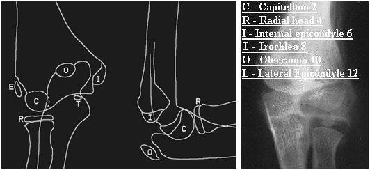
child’s development and can quite easily be mistaken for bony injury. Remember that the smooth rounded appearance of a centre of ossification does not often mimic the typically sharp edges of a new fracture fragment, but even saying this, distinction can be very difficult. If you have the CRITOL (or CRITOE) acronym in your mind, it will help you to interpret the xray with a sensible approach. The image below highlights each of the six ossification centres with the typical age of the child when each centre appears:
Note: Lateral / External epicondyle are interchangeable terms, giving either CRITOL or CRITOE
Work through in order the centres of ossification – each may appear at slightly different ages in different children, but the sequence in which they
appear should always be C,R,I,T,O,L. For example in a 6 year old you would not expect the lateral condyle to have appeared yet, so if there is a bony fragment at that site, it is suggestive of a fracture.
Supracondylar Fractures:
The supracondylar fracture (of the distal humerus) is the most common upper limb fracture in young school age children. It presents almost always as
a FOOSH (fall on the outstretched hand). They are likely to be in a lot of pain, and may need strong analgesia and immobilisation (with a splint or
sling) before assessment is possible. (A good example of a strong, rapid acting, well tolerated analgesic that can be used in the Emergency Department is intranasal diamorhpine, which is made up with saline into a small volume of fluid and then given as nose drops with a syringe.)
You may well see bruising and swelling around the elbow, with tenderness around the distal humerus, but remember to check the whole limb including
clavicle (and the whole of their body if the mechanism of injury could have caused other serious injuries!)
The elbow contains numerous important neurovascular structures – your assessment must document presence of radial pulse, capillary refill time in
fingertips and function of ulnar, median and radial nerves (not possible formally in younger children, so watch to see if they will hold a toy or parents hand
and be suspicious if the xray shows a displaced fracture). Any concerns about neurovascular compromise require the fracture to be urgently reduced, either by the Emergency Department team or referral to Orthopaedics. This initial reduction is to relieve mechanical pressure of displaced bony fragments on the
neurovascular structures and will not provide the stability required for neatly aligned healing, so a trip to theatre for fixation will happen soon after.
A common classification system for supracondylar fractures is the Gartland classification. In this there are three categories of supracondylar fractures based on their radiological appearance on the lateral elbow view:
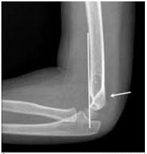
1: undisplaced – recognised as clinical suspicion plus xray evidence of fluid in the elbow joint elbow (as demonstrated by a raised anterior fat pad or
the presence of a posterior fat pad) and possibly loss of anterior humeral line (normal is when line along anterior humerus intersects middle third of
capitellum)
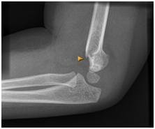
2. visible fracture which is hinging on the posterior edge
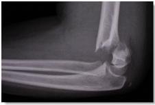
3. totally displaced
Type 1 can usually be managed conservatively with an above elbow cast and sling, whilst 2 and 3 require operative fixation.
All should be followed up by Orthopaedics in fracture clinic – typical progress is evidence of union at 4 weeks, and then the patient is encouraged to
gradually do away with the sling, to allow mobilisation without too much discomfort.
A rare but debilitating late complication of supracondylar fractures is Volkmann’s ischaemic contracture (when the brachial artery is damaged and months later the patient develops clawing of the thumb and fingers and forearm wasting).
Minor injuries introduction
Minor injuries Series: Episode 1 with thanks to Dr Jessica Spedding, PEM trainee, Royal London Hospital, UK
Introduction to minor injuries:
Minor injuries in children are common and mostly self limiting soft tissue injuries that heal with time. Some injuries are particular to paediatrics (pulled elbow) and others are simply much more common in children than adults (supracondylar fracture). Another consideration specific to children is consideration of growth plate involvement, which if does not heal in a good position could lead to asymmetry and growth problems. Injuries involving the growth plate are graded as Salter-Harris 1,2,3,4, or 5 and they will be discussed in more detail in a future episode of this minor injuries series.
Your assessment:
You need a systematic approach that assesses for important injuries that need specific management. Your
assessment must always include consideration of non accidental injury (NAI). A sensible approach would include:
– Is the mechanism of injury described consistent with the injury sustained?
– Has the child reached the appropriate stage of development to have sustained the injury in the way described?
– Is there any delay in presentation?
– Has the child (or siblings) presented numerous times before with injuries?
– There is an excellent set of pamphlets that give evidence based guidance on when injuries point to abuse – go to www.core-info.co.uk or look out for the summaries on Paediatric Pearls
Upper limb injuries:
You may have come across the acronym FOOSH. This is a Fall On the Out-Stretched Hand. This mechanism is the natural response to a fall – in order to protect our head and trunk, the reflex is to put our arms out to break our fall. This mechanism causes a number of different injuries, each more prevalent in different age groups (but common in other age groups too). Roughly speaking these could be sequenced as follows:
Age 1-3: distal radius fracture (usually greenstick or torus) or middle third clavicle fracture
Age 4-8: supracondylar fracture (varying degrees of severity, some of which require operative fixation)
Age 9-adulthood: distal radius fracture or scaphoid fracture
However one must still examine unclothed the whole limb to be sure that all sites of injury have been located. In the upper limb this would be from
fingers to shoulder, clavicle and possibly neck, in the lower limb this would be from toes to hips but also checking the pelvis and lower spine.
The first chapter in this series looks at a common elbow problem:
Pulled elbow: (see also http://www.paediatricpearls.co.uk/2012/02/pulled-elbow/)
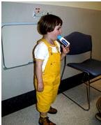 Proper name – radial head subluxation, mechanism is usually a sudden pulling of the child by their hand (such as to stop them running into the road or swinging a child in play), child presents having cried initially, but soon settles but refuses to use the arm, holding it slightly flexed at the elbow with the arm by their side. When you go to assess them they have no swelling or bruising or distal neurovascular compromise, but are very apprehensive about you trying to bend or pronate/supinate the elbow. In up to half of cases there may not be a “pull” mechanism in which case be more cautious in assuming the diagnosis. Don’t forget a clavicle fracture may present this way. If you feel sure the diagnosis is pulled elbow, attempt a reduction as follows:
 Hold their hand as though you were going to shake hands, with your other hand gently cupping underneath the elbow, with elbow partially flexed, then firmly pronate (rotate to palm up position). You should feel the clunk of a reduction, but if not, try a firm supination (back to palm down position). Ideally do this half an hour after some analgesia. If you do not feel a clunk it is probably not reduced but either way stop after two attempts, and then allow the child to be somewhere relaxing and ask their parent to let you know if they start playing – if reduced most will soon realise the pain with movement has gone and start playing normally within a few minutes. If not reassess and consider a differential diagnosis which may include referral for xray.
August 2012 PDF digest
August’s PDF only has 4 text boxes but with lots of information crammed into them and extra on the blog. A great looking PDF on poisoning in children from one of our registrars, an article on stammering from another working with a speech and language therapist and an update on BTS pneumonia guidelines just in time for the winter. Also a feature on Cardiff’s core info safeguarding work on the evidence behind different types of fractures. Do leave comments…
