Scabies this month with a beautiful picture of plantar lesions in a child. Updated NICE head injuries, antipyretics (or not) for febrile convulsions, child trafficking and the last in the sleep series. Do leave comments below.
Tag Archives: head injury
August 2013 PDF published
Thermal injuries from a safeguarding point of view this month, updated fever guidelines, quarantine periods for infectious diseases, house dust mite allergy and facial injuries this month as the last in the minor injuries series. Do leave comments below.
July 2013 PDF
Neglect and emotional abuse is the safeguarding topic this month. ED advice on the management of minor head injuries, a report from BPSU in hypocalcaemic fits secondary to vitamin D deficiency, the new UK immunisation poster and a bit on crying babies. Hope you find it all helpful. Comments welcome below
child abuse and head injuries
This summarises the Core-info leaflet on head and spinal injuries in children. Full details are available at www.core-info.cardiff.ac.uk.
**PLEASE REFER ALL SUSPECTED INFLICTED HEAD AND SPINAL INJURIES TO PAEDIATRICS **
Inflicted head injuries
- can arise from shaking and/or impact
- occurs most commonly in the under 2’s
- are the leading cause of death among children who have been abused
- survivors may have significant long term disabilities
- must be treated promptly to minimise long term consequences
- victims often have been subject to previous physical abuse
Signs of inflicted head injury
- may be obvious eg. loss of consciousness, fitting, paralysis, irritability
- can be more subtle eg. poor feeding, excessive crying, increasing OFC
- particular features include retinal haemorrhages, rib fractures, bruising to the head and/or neck and apnoeas
- also look for other injuries including bites, fractures, oral injuries
If inflicted head injury is suspected
- a CT head, skull X-ray and/or MRI brain should be performed
- neuro-imaging findings include subdural haemorrhages +/- subarachnoid haemorrhages (extradural haemorrhages are
more common in non-inflicted injuries) - needs thorough examination including ophthalmology and skeletal survey
- co-existing spinal injuries should be considered
- any child with an unexplained brain injury need a full investigation eg. for metabolic and haematological conditions, before a diagnosis of abuse can be made
The following diagram comes from http://www.primary-surgery.org:
These CT images are from http://www.hawaii.edu/medicine/pediatrics/pemxray/v5c07.html:
EXTRADURAL (or epidural) haematoma
SUBDURAL haemorrhages in a 4 month old
SUBARACHNOID haemorrhage in a 14 month old
Neuro-imaging for inflicted brain injury should be performed in
- any infant with abusive injuries
- any child with abusive injuries and signs and symptoms of brain injury
Inflicted spinal injuries
- come in 2 categories : neck injuries, and chest or lower back injuries
- neck injuries are most common under 4 months
- neck injuries are often associated with brain injury and/or retinal haemorrhages
- chest or lower back injuries are most common in older toddlers over 9 months
- if a spinal fracture is seen on X-ray or a spinal cord injury is suspected, an MRI should be performed
Fractures in child abuse
Metaphyseal fractures, also known as a bucket handle, chip or corner fracture, occur at the growing end of the bone and only in children. Recent fractures are very difficult to see on x-ray and they are often not associated with any clinical sign of soft tissue swelling or bruising. They may become more obvious radiographically after 11 to 14 days. They are thought to happen when the baby has been pulled or swung violently and the relatively weaker growing point of the bone breaks. They have been noted to occur accidentally following birth injuries, following serial casting of talipes or as a consequence of appropriate physiotherapy to newborn babies. (Source: Core-info leaflet)
The picture of a metaphyseal fracture of an infant’s wrist below comes from a 2000 paper on the orthopaedic aspects of child abuse by Kocher et al and published in the Journal of the American Academy of Orthopaedic Surgeons (http://www.jaaos.org/content/8/1/10.abstract):
A spiral fracture refers to the direction in which the bone is fractured. It implies that there has been a twisting force to cause the fracture. Spiral fractures can also occur accidentally in the femur once the child is walking. (Source: Core-info leaflet)
The picture below of a spiral fracture of the femur in a 2 month old comes from http://www.hawaii.edu/medicine/pediatrics/pemxray/v6c02.html (same website has a wealth of other paediatric radiological images on it if you are interested):
A supracondylar fracture is one in the upper arm immediately above the elbow and is highly suggestive of accidental injury. The picture below comes from http://www.kidsfractures.com/, a site put together by 2 American orthopaedic surgeons who say their aim was to make parents’ experience of having a child with a broken bone a little less traumatic but I think much of the language and many of the pictures are more suited to a medical audience.
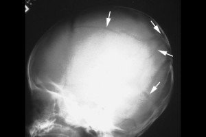
A simple linear skull fracture is a break in a cranial bone resembling a thin line, without splintering, depression, or distortion of bone. They are equally prevalent in NAI and in accidental injury. The picture below is of a linear right parietal skull fracture and comes from a Rumanian educational website, http://www.medandlife.ro/medandlife602.html.
This compares with a complex skull fracture which is variously defined as:
• a depressed fracture (where the skull is pushed in)
• two or more fractures of the skull
• fractures that cross the sutures (natural joining edges of skull bones) or those that are widening. (Source: Core-info leaflet)
The picture below of a complex skull fracture is from http://www.childabuseconsulting.com/child-abuse-fractures.html which houses other not-very-subtle images of non-accidental burns and bite marks as well.
Rib fractures in infants, particularly posterior ribs, with no history of major trauma are suspicious. The picture below is taken from http://www.learningradiology.com/notes/bonenotes/childabusepage.htm and shows multiple rib fractures with callous formation, the ones of the left 2nd and 6th posterior ribs being the easiest to identify:
December 2011. Happy Christmas!
December 2011 has snippets of information on torticollis (backed up with lots more information on the website), unconscious children, alkaline phosphatase and a link to the Map of Medicine’s recent algorithm for cough in children. Also some pointers for your safeguarding training needs. Download it here.
January ED version of Paediatric Pearls newsletter
This month’s emergency department version of Paediatric Pearls has information on the NICE guideline on head injury, what to do if you find a child has an undescended testis and some pointers to sites on asthma inhalers. Download the January 2011 ED version here.
January GP edition here!
January reminds us all of the NICE guideline on head injury and specifically when a child is supposed to be referred for a CT. We continue our 6-8 week baby check series with information on undescended testes. There are also links to agreed blood test reference ranges and resources to help with the identification of asthma inhalers. Download January 2011 GP PDF here.
Head injury
This month I have featured the 2007 NICE guideline on head injury. This is because I was looking through it recently trying to find out how long we should be observing children with minor head injuries for in A and E ie. those who do not qualify for a CT. I was also interested to find out whether we should treat babies, whose fontanelles are still open, differently. Would it take longer for the signs of intracranial pressure to become obvious in them? Anyway the guideline answers neither of those questions…
I found a couple of recent Canadian papers on the need for CT scanning in children with minor head injury. I think many of us are concerned that we are doing too many CTs on children as a result of the NICE guideline. The radiation dose is not insignificant and some of the children have to be sedated for the investigation which is an added risk too. Maguire et al (Should a head injured child receive a head CT scan? A systematic review of clinical prediction rules. Pediatrics 2009;124(1):e145 – e154) say in the introduction to their paper that up to 70% of children presenting to the ED in the USA or Canada with a head injury get a CT scan and 70% to 98% of them are normal. A more recent attempt at a clinical decision tool for assessing the need for CT has been written up by the Canadian head injury study group: Osmond MH et al. CATCH: a clinical decision rule for the use of computed tomography in children with minor head injury. CMAJ 2010;182(4):341-348. There is a nice summary of and comment on this paper at www.medscape.com/viewarticle/579598.
The Scottish intercollegiate network (SIGN) put together a very similar guideline on head injury in 2009. Their patient information leaflet is, in my view, infinitely better than NICE’s. Have a look at their documents and downloads at http://www.sign.ac.uk/guidelines/fulltext/110/index.html


