Well the BMJ produces 2 journals in one in August so why can’t I? All the topics featured this month are relevant for both GPs and ED doctors – for once – so you have a joint newsletter. I have covered headache this month, Vitamin D (by popular request) and we have started the “Feeding” series requested by my ED senior colleagues. It seems appropriate to have covered breastfeeding first. Do leave comments below.
Tag Archives: newborns
Common breastfeeding problems
My ED consultant colleagues requested that we run a series on “feeding issues” in Paediatric Pearls as it forms a part of the ED trainees curriculum and is a common subject to come up in conversation with parents in the ED. It seems appropriate to begin the series with an article on breastfeeding problems put together by our breastfeeding counsellor, Jo Naylor, and one of the current paediatric trainees, Dr Sarah Prentice. Their full article is downloadable here and I have reproduced some nuggets in this month’s Paediatric Pearls newsletter and below.
| Breastfeeding adequately? | Inadequate milk intake? |
| feeding every 2 – 5 hours for 20 – 40 minutes | infrequent feeds |
| 3-4 wet nappies and changing stool by day 3 | continued urates and/or meconium after day 3 |
| pain free breastfeeding | painful feeds, ineffective sucking |
| weight loss < 10% | weight loss > 10% |
| baby settled between feeds | fretful, hungry baby |
Reminder: handout of local breastfeeding drop-in groups available here.
I intend to cover the following topics over the next few months (some of which have actually already been touched on in previous months): vitamin supplementation, formula milk, gastro-oesophageal reflux, starting solids, allergy, fussy eating, food refusal, dentition and use of bottles, healthy eating, obesity, eating disorders. Please do leave requests for other topics below.
Inguinal hernias
with thanks to Dr Jemma Say for putting the following information together:
Inguinal Hernias
An indirect inguinal hernia is a protrusion of abdominal contents into the inguinoscrotal or labial canal via an open deep inguinal ring due to the failure of obliteration of the processus vaginalis.
In fetal life the descent of the testis into the inguinal canal and scrotum is preceded by a small pouch of peritoneum; the processus vaginalis. After birth this peritoneal communication is obliterated, failure to do so results in either a hydrocele or hernia, depending on the degree of fusion.
Indirect hernias are more commonly seen in a paediatric population, as opposed to direct inguinal hernias in adult patients, where the musculature is weak and abdominal contents protrude through the wall of the inguinal canal.
Epidemiology
The incidence is 1-2%, occurring 9 times more commonly in males. The majority are found on the right (60%), 15% are bilateral, more commonly with a family history. Presentation is most frequently in infancy.
Increased Incidence
- Preterm infants (10-30%)
- Abdominal wall defects (e.g. prune belly syndrome)
- Connective tissue disorders (e.g. Ehlers Danlos syndrome)
- Chronic respiratory disease
- Undescended testes
- Increased intraabdominal pressure
The diagnosis is clinical, although USS can play a role in older children with indeterminate pain. Surgery is indicated for all paediatric patients with inguinal hernia.
The risks of not performing surgery include bowel incarceration or necrosis, and testicular or ovarian compromise and necrosis. This risk is greatest in early infancy; premature infants have an incarceration risk of up to 30%, and therefore often warrant treatment prior to discharge. Some surgeons keep under close review for a few weeks post discharge so that these still very small babies put on a bit of weight before the operation.
If a patient presents with incarceration, an attempt at reduction should be made and urgent surgery is required, as the risk of reincarceration is as high as 15% if surgery is delayed more than 5 days.
Referral Pathway
All inguinal hernias should be referred, paediatric patients >1 year can be referred to Mr Brearley at Whipps Cross while those <1 should be referred to the Royal London Hospital. Surgery involves either open or laparoscopic techniques (tranperitoneal or preperitoneal approaches). The majority are performed as an outpatient with normal activity resuming within 48 hours.
References
IPEG guidelines for Inguinal Hernia and Hydrocele, Nov 2009. http://www.ipeg.org/education/guidelines/hernia.html
Ashcraft’s Paediatric Surgery, Holcomb G W, Murphy. J P
ABC of General Paediatric Surgery: Inguinal hernia, hydrocele and the undescended testis: BMJ 1996 312:564
Patient Information Leaflets
http://www.patient.co.uk/doctor/Inguinal-Hernias.htm
http://www.bch.nhs.uk/acrobat/PDF%20for%20Web/Inguinal%20Hernia%20Repair.pdf
Video information
Distinguishing indirect and direct inguinal hernia
http://www.youtube.com/watch?v=wAzXSqGybvE
Indirect inguinal hernia repair
http://www.medicalvideos.us/play.php?vid=1108
Contraindications to breastfeeding
I was encouraging a mother to breastfeed the other day when she asked if I was sure that was OK with her condition. Her baby is asymptomatic on a 10 day iv course of penicillin for presumed inadequately treated maternal syphilis. I wobbled momentarily and the junior doctor and I went away to look it up. It is OK apparently as long as the mother does not have syphilitic lesions around her nipples. Take a look at http://pedclerk.bsd.uchicago.edu/page/breastfeeding which is an American teaching site and has a nice summary of when you can and can’t breastfeed. http://toxnet.nlm.nih.gov/cgi-bin/sis/htmlgen?LACT is an American database of the evidence on the safety of various medicines when breastfeeding.
March Paediatric Pearls for GPs
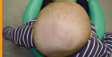
The March 2011 version is now published. I have covered the new NICE guideline on food allergy which I think you will all find helpful and provided a link to the Allergy Academy which runs some really excellent course on all aspects of allergy in children. We continue with the 6 week check series with some information and pictures on fontanelles, craniosynostosis and positional plagiocephaly. There’s a bit on how to get foreign bodies out of noses. Do leave comments below.
FONTANELLES AND HEAD CIRCUMFERENCE AT SIX WEEK CHECK
with thanks to Dr Harriet Clompus.
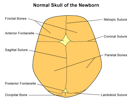
Assessment of fontanelles is an important part of the six week check. Large fontanelles may indicate a problem in bone ossification or hydrocephaly, while a fused anterior fontanelle can indicate craniosynostosis. These need to be referred to paediatric outpatients. Always remember that anterior fontanelle size is very variable (1-4.7 cm in any direction) and always needs to be assessed in context of baby’s head circumference.
A sunken fontanelle indicates dehydration, while a bulging fontanelle indicates raised intracranial pressure (but can be non-pathological – vomiting, crying, coughing – so assess when baby settled!). These can be discussed with paediatric registrar on-call.
1) The anterior fontanelle is diamond shaped, 1-4.7 cm in any direction at birth (black infants larger than white) and can widen in first 2 months of life. Median age of closure is 14 months (4 – 24 months)
2) The posterior fontanelle is triangular and is less than 1 cm. It closes by 6 – 12 weeks.
3) The size of the fontanelles should always be assessed in conjunction with the head circumference.
- Macrocephaly – familial, hydrocephaly or skeletal disorders such as achondroplasia.
- Microcephaly – familial, congenital infections, fetal alcohol syndrome, trisomies
4) The quality of the fontanelle should always be assessed.
- Soft fontanelle – normal
- Bulging fontanelle – raised intracranial pressure (hydrocephalus, meningitis/encephalitis) . NB can be non-pathological in crying, coughing or vomiting infant.
- Sunken fontanelle – dehydration
1) WIDENED FONTANELLES: think of…
Achondroplasia
Downs
Hydrocephalus
IUGR
Prematurity
Congenital Rubella
Neonatal Hypothyroidism (3rd fontanelle)
Osteogenesis Imperfecta
Malnutrition
Rickets/Osteomalacia
Rickets – Think of rickets in darker skinned, breast fed babies, especially if mothers are veiled. Infants will often have sweating on the head. If widened sutures are found check neonatal blood spot for hypothyroidism and refer to outpatients.
Hydrocephalus – can have widened, bulging fontanelles in conjunction with a large head.
2) PREMATURE FUSION OF FONTANELLES AND CRANIOSYNOSTOSIS
Closure of anterior fontanelle by six weeks always pathological (NB by 3 months 1% of normal infants will have a closed anterior fontanelle).
Must always assess in conjunction with head circumference – early fusion associated with microcephaly (and less commonly, macrocephaly).
Craniosyntosis is premature closure of cranial suture(s) with skull growth restriction perpendicular to fused suture and compensatory skull overgrowth in unrestricted areas. Presents with ridging (always pathological beyond one week of life) and abnormal skull shape (usually later than six weeks).
There is a nice background overview (with useful diagrams) to craniosynostosis at http://www.cincinnatichildrens.org/health/info/neurology/diagnose/craniosynostosis.htm.
Primary craniosynostosis is due to abnormal ossification of one or more sutures. Simple – premature fusion of one suture, complex – premature fusion of multiple sutures. Causes include rickets, hyperparathyroidism, hyperthyroidism , idiopathic and genetic causes such as Aperts.
Secondary craniosynostosis is caused by premature closure of ALL sutures due to lack of primary brain growth. If you find a child with premature closure of fontanelles or over-riding sutures at six week check you should refer to paediatric outpatients. NB Plagiocephaly (flat occiput) is a non-pathological deformation due to ‘back to sleep’ position – no action required. It presents with ear on flattened side presenting anteriorly. Parallelogram shaped head (as opposed to lambdoid suture craniosynostosis trapezoid shaped)
The following articles give lots of information on fontanelles and/or sutures. The Fuloria article is very thorough and although it focuses on neonatal examination, most of it is still relevant for the six week check.
1) The Abnormal Fontanel, J KIESLER et al Am Fam Physician. 2003 Jun 15;67(12):2547-2552. http://www.aafp.org/afp/2003/0615/.html (figure 2 taken from abnormal fontanel)
2)The Newborn Examination: Part I. Emergencies and Common Abnormalities Involving the Skin, Head, Neck, Chest, and Respiratory and Cardiovascular Systems, Fuloria et al, Am Fam Physician. 2002 Jan 1;65(1):61-69. http://www.aafp.org/afp/2002/0101/p61.html
3) http://www.nice.org/CG037quickrefguide
4) http://www.patient.co.uk/doctor/Examination-of-the-Neonate.htm
5) Craniosynostosis, P Raj et al, emedicine jul 2010 Craniosynostosis : eMedicine Neurology
Neurological examination in babies
I have been looking at information on primitive reflexes as I was asked by a GP whether it was significant if he could not elicit a Moro reflex at the newborn check. Wikipedia has a nice description of the Moro reflex: “it may be observed in incomplete form in premature birth after the 28th week of gestation, and is usually present in complete form by week 34 (third trimester). It is normally present in all infants/newborns up to 4 or 5 months of age, and its absence indicates a profound disorder of the motor system. An absent or inadequate Moro response on one side is found in infants with hemiplegia, brachial plexus palsy, or a fractured clavicle”.
You need to make sure you are eliciting it correctly first though. I have found a great site from Utah university with little video clips of aspects of a normal and abnormal neurological examination of a 5 day old. Take a look at http://library.med.utah.edu/pedineurologicexam/html/newborn_n.html and watch the 2 Moro examples carefully. Did you know the hands have to come together in the mid-line at the end?
GP version of February 2011’s Paediatric Pearls
GP February 2011 reminds us all of the NICE guideline on Attention Deficit and Hyperactivity Disorder. We continue our 6-8 week baby check series with information on the absent red reflex and go back to our “from the literature” box to discuss snoring and obstructive sleep apnoea (OSA). We have relaunched our prolonged jaundice guideline. Please leave comments and questions below.
Checking the red reflexes
6 week check series – The Absent Red Reflex – with thanks to Dr Sarah Prentice
Importance of red reflex examination at the 6 week check
Early detection of potentially sight and life-threatening eye disease. Due to the early and time-limited plasticity and development of the eye, any blockage of light to the retina interferes with development of optic neural pathways and can have profound effects on later vision.
Pathology
Cataracts
Retinoblastoma
High Refractive errors
Vitreal haemorrhage/opacity
Corneal scaring (e.g. ocular toxocariasis)
Retinal tear
Retinopathy of prematurity
Persistence of the tunica vasculosa lentis/Persistent hyperplastic primary vitreous (1)
The Examination
Darkened room
Ophthalmoscope on +3 dioptres
Hold 1 foot away
Red reflexes can only be described as normal if they are:
Equal in colour, intensity and clarity with no opacities or white spots (2)
Handy hints
For the child that won’t open his/her eyes: try picking/sitting them up or rocking them from sitting to lying. Having a parent hold them on their shoulder (as if winding them) and looking from behind often works. A feeding child will often open his/her eyes, although breast feeding then makes looking in the eyes logistically tricky.
Children with darker skin tones may have pale retina. If retinal vessels can be seen and followed to the disc and the reflex is equal bilaterally then this is reassuring. Comparison with parents’ red reflexes may also help.
Management:
Normal: No further follow-up. Will have routine ophthalmology review by school nurse/orthoptist in pre-school years. (5)
Unable to see red-reflexes or unsure: Referral to paediatric ophthalmology primary care clinic (if available)
Absent red reflex: Urgent referral to paediatric ophthalmologists (should be seen in less than 2 weeks)
Family history of neonatal eye disease e.g. retinoblastoma, congenital cataracts: Routine referral to paediatric ophthalmologists.
Low birth-weight/premature infants (at high risk of retinopathy of prematurity): Should have had ROP screening and follow-up arranged as necessary by neonatal unit.
References and resources
- 1. Robertson’s Textbook of Neonatology. Fourth Edition. 2005. Edited by Janet M. Rennie.
- 2. American Academy of Pediatrics Policy Statement. Red Reflex Examination in Infants PEDIATRICS Vol. 109 No. 5 May 2002
- 3. Red Reflex Examination in Neonates, Infants, and Children. PEDIATRICS Vol. 122 No. 6 December 2008, pp. 1401-1404 (doi:10.1542/peds.2008-2624)
- 4. www.eyesite.ca/7modules/Module5/html/Mod5Sec1.html – Good pictures of cataracts, retinoblastoma and glaucoma
- 5. http://www.patient.co.uk/doctor/Vision-Testing-and-Screening-in-Young-Children.htm
- 6. http://www.bartsandthelondon.nhs.uk/docs/poster_red_reflex_print.pdf Poster of red reflexes and referral pathway from Bart’s and The London.
December 2010 PDF digest for GPs now published!
December’s Paediatric Pearls (GP edition) reminds us all of the NICE guideline on antibiotic prescribing in respiratory tract infections. I would like to do a bit more of the “delayed prescribing” in the Emergency Department but it would require either the family coming back (ie. a “no antibiotic” policy really) or their putting a bottle of amoxicillin in their fridge and potentially not using it as we give out the actual antibiotic in A and E, not prescriptions. We’ve also featured a couple of papers showing that chest x-rays add very little to the management of a child with a respiratory illness which I think most GPs know but it doesn’t harm to remind trainees still in the hospital that, just because the radiology department is at the end of the corridor, it doesn’t mean you have to use it! We continue our 6-8 week baby check series with information on sacral dimples and I have also put in a couple of websites with sensible, empathetic information and advice on school refusal. The beginning of term is stressful for children who find it hard to go to school and parents may find these sites helpful when trying to understand why their child is behaving in that way. Happy New Year to you all!

