Category Archives: For General Practitioners

Disordered puberty neatly explained

With thanks to Dr Amy Rogers for unravelling endocrinology for the better understanding of all non-endocrinologists.  All girls under 8 and boys under 9 with signs of puberty should be referred to a paediatrician and you could leave it at that.  But for those who want to know a bit more about it (or check up on what we do about it…) read on!

The British Society of Paediatric Endocrinology and Diabetes (BPSED) is recognised by the Royal College of Paediatrics and Child Health (RCPCH) as the society responsible for this field of paediatric medicine. It is currently chaired by Professor Dattani of Great Ormond Street Hospital and represents the only U.K. society responsible for governing the training of doctors in paediatric endocrinology and diabetes and actively supporting the ongoing training and education of allied healthcare professionals in this specialist area. Lots of resources available at www.bsped.org.uk


The hormones of puberty: Hypothalamic-Pituitary-Gonadal Axis:

Hormones of puberty
Hormones of puberty


(http://www.ohioshaolindo.com/China%27s%20Arts/image016.jpg)


Start of puberty in girls = palpable breast bud (B2)

Start of puberty in boys = testicular vol >3.5ml


Androgens promote other secondary sexual characteristics: smelly feet, acne, body odour and mood swings!


Timing of puberty is dependent on socioeconomic status, nutritional and genetic factors.


In the UK early puberty = <8years girls, <9 years boys. Interpret in conjunction with family history (age of maternal puberty), ethnicity (Afro-Caribbean or mixed race = commoner to have early menarche), BMI (overweight = associated with early puberty), social factors (adoption and lower SEC = early puberty) and past medical events. Approx 10% girls achieve menarche whilst still in primary school.


RED FLAG: ARRESTED PUBERTY = SERIOUS PATHOLOGY


Pubertal development may be:


1)       Concordant (following the normal pattern), i.e. breast buds, pubic hair then menses/increased testicular vol, pubic hair then penile enlargement).

OR


2)       Discordant, e.g. progressive breast enlargement with no pubic hair, or penile enlargement with small testicles. Suggests over activity of sex hormone (oestrogen/testosterone) production in the periphery, i.e. adrenals, gonads (ovaries/testes) or tumours.


Causes of precocious puberty


Central precocious puberty (gonadotrophin dependent)

  • Idiopathic = Most common cause in girls (10 times more common than boys). Slowly progressing (breast and pubic hair growth, modest growth spurt, bone age advanced <1yr, few changes on pelvic USS) or more aggressive (height velocity greater, bone age advanced >1 yr). Ovarian and uterine enlargement (>2ml) on USS. Pubertal response to stimulation test. Boys: testicular enlargement, virilisation, pubertal response to stimulation test.


  • Tumour = Second most common cause in both sexes (at least half of all boys presenting with central precocious puberty), typically a hypothalamic hamartoma


  • Optic glioma (NF)
  • Longstanding/severe peripheral secretion
  • Abnormal brain (hydrocephalus, septo-optic dysplasia)
  • CNS damage (infection/trauma/ low dose irradiation)
  • Adoption
  • HCG production (CNS or peripheral tumours)
  • Hypothyroidism


Peripheral precocious puberty (gonadotrophin independent): peripheral oestrogen

Thelarche

Ovary tumour or cyst (including McCune-Albright syndrome and massive ovarian oedema)

Drug/dietary sources

Testicle/liver tumour


Peripheral precocious puberty (gonadotrophin independent): peripheral androgen

Adrenarche

Atypical congenital adrenal hyperplasia (CAH)

Adrenal tumour (including Cushing’s)

Testicular tumour

Testotoxicosis (male limited family history) and McCune-Albright syndrome (irregular café-au-lait patches, bony changes on xray and ovarian cysts (that may be huge).


Investigation


Examination alone is often enough to determine if this is “true” puberty or just early thelarche (breast development) or pubarche (body hair), especially if combined with bone age.


  • X-ray right wrist (bone age)
  • Pelvic and abdominal USS (looking for tumours, cysts, size and position of gonads/internal genitalia)


Definitive test for central precocious puberty = GnRH stimulation test:


1)       Measure LH, FSH, oestrogen/testosterone at base-line.

2)       Give GnRH and monitor serial response of LH and FSH (20mins and 60mins)


Interpretation:


20mins 60mins
LH/FSH Central precocious puberty
LH/FSH Peripheral precocious puberty


MRI hypothalamus, pituitary and brain in aggressive forms of early puberty, in girls less than 6 years of age, any child with neurological signs, and all boys.


Other tests to consider:

  • TFTs (elevated TSH in hypothyroidism can mimic FSH, inducing early testicular and ovarian enlargement)
  • Tumour markers (HCG can be produced from pineal, hepatic and testicular tumours) and alpha fetoprotein will be raised but LH/FSH will be low and non-stimulatable.
  • Testosterone, oestrogen, morning LH/FSH


Treatment

1)       Early thelarche or pubarche: None. However, if pubarche associated with being overweight in girls, important to control weight, otherwise at increased risk of polycystic ovary syndrome (PCOS) and Type 2 diabetes later in life.


2)       Supportive, let nature take its course: coping with periods, behavioural and cosmetic changes. School need to be aware.


3)       Triptorelin or Goserelin injection = long acting GnRH analogues (given every 4-12 weeks depending on preparation used and body’s response to it). Will slow down or stop development. Continued until child’s peers are entering puberty, typically aged 10-11years. GH in addition, may result in improved final height, in girls.


Additional notes on discordant sexual development:


Thelarche: commonly present from infancy, non-progressive, may be unilateral. No treatment required.


Ovarian (and adrenal, testicle or liver) tumours secreting oestrogen are rare and often present with a palpable mass and prominent breast development with no other signs of puberty.


Thelarche-like symptoms are produced from oestrogen containing medications.

Adrenal androgens are the commonest cause of early virilisation leading to sexual hair growth, body odour, acne, greasy hair and mood swings.


Adrenarche = normal maturation of the adrenal glands leading to enhanced secretion of the androgen DHEA. Usually co-existent with the onset of normal puberty and contributes most of the androgenic component of puberty in young females. Premature adrenarche may be familial, spontaneous, or with an ill-understood association with hydrocephalus. No treatment required. A proportion of girls affected will proceed to PCOS, especially if they gain excessive weight.


Atypical CAH (mild, non-salt wasting type) mimics adrenarche and can be easily differentiated by a urinary steroid profile or a short Synacthen test and measurement of 17 alpha hydroxyprogesterone. A pubertal response to stimulation testing will require treatment with both hydrocortisone and GnRH agonists to achieve a reasonable final height.


Non-iatrogenic Cushing’s syndrome tends to be accompanied by excess adrenal androgen secretion and hirsuitism.


Isolated premature menarche is relatively common disorder of unknown aetiology. USS demonstrates prepubertal uterus with no endometrial lining between bleeds. Differential diagnosis includes rare local lesions, e.g. sarcoma, abuse and vaginal foreign body.


Mild, transient breast enlargement occurs in approx 50% boys in early puberty, but severe persistent gynaecomastia is increasingly common, possibly secondary to nutritional excess or environmental chemicals. Usually accompanies early puberty but can be pre-pubertal. Investigations for ectopic oestrogen secretion, karyotype, liver and thyroid function are usually normal. If present for >18m, may require surgical removal. If diagnosed early, treatment with anti-oestrogen medication such as anastrozole may have some benefit.

Paediatric Pearls for February 2012

Click here for this month’s PDF digest!  It ‘s quite hard providing a balance of information for GPs and ED juniors now that I am only doing the one newsletter.  I think we’ve succeeded this month with neurodevelopmental milestones in Down’s syndrome and essential tremor aimed mainly at GPs and pulled elbow, anaphylaxis and the FEAST study aimed more towards the emergency medicine practitioners.  Many thanks to my colleagues who have contributed this month.  The FEAST video makes fascinating and inspiring watching for any health professional, regardless of specialty.  Do leave comments, questions, suggestions!

NICE on anaphylaxis

With thanks to my colleague, Dr Su Li, for summarising this 2011 NICE guideline for Paediatric Pearls.

Anaphylaxis: assessment to confirm an anaphylactic episode and the decision to refer after emergency treatment for a suspected anaphylactic episode

December 2011

www.nice.org.uk/cg134

Anaphylaxis is a severe, life-threatening, generalised hypersensitivity reaction involving

  • the airway (pharyngeal or laryngeal oedema) and/or
  • breathing (bronchospasm, tachypnoea) and/or
  • circulation (hypotension, tachycardia).


There can often be skin and mucosal changes. Patients presenting with these signs and symptoms should be diagnosed as having ‘suspected anaphylaxis’.

Anaphylaxis may be an allergic response that is

  • immunologically IgE mediated (foods, venoms, drugs, latex) or
  • non-immunologically mediated or
  • idiopathic (significant clinical effects with no obvious cause).


This guideline does not make any drug recommendations. These can be found at http://www.resus.org.uk/pages/reaction.pdf.

Patient Centred Care

  • Treatment and care should take into account patient’s needs and preferences
  • Patients should have the opportunity to make informed decisions about their care and treatment, in partnership with health care professionals
  • Good communication between healthcare professionals and patients is essential
  • Families and carers should be given the information and support they need
  • Care of young people in transition between paediatric and adult services should be planned and managed according to the best practice guidance described in ‘Transition: getting it right for young people


Recommendations

  • Document acute clinical features of the suspected anaphylaxis
  • Record the time of onset
  • Record the circumstances immediately before the onset of symptoms to help identify possible triggers


  • Consider taking blood samples for mast cell tryptase if reaction is thought to be immunologically mediated or idiopathic
    • First sample as soon after emergency treatment given
    • Second sample 1-2 hours (no more than 4 hours) from onset of symptoms
    • A further sample may be required at follow up with the allergy specialist to measure baseline mast cell tryptase


  • Children who have had emergency treatment should be admitted to hospital under the care of the paediatric team.  The resus council suggests observing the child for a pragmatic (no evidence yet) 6 hours because of the risk of a biphasic reaction.
  • Offer the child/parents a referral to an allergy specialist (see www.bsaci.org for registered allergy clinics)
  • Offer the child/parents an adrenaline injector in the interim period whilst waiting for a specialist appointment


  • Before discharge, offer the child/parents
    • Information about anaphylaxis (signs, symptoms, risk of recurrence of symptoms (biphasic reaction)).  Parent information leaflet here.
    • Information about what to do if a reaction occurs (use adrenaline injector, call emergency services)
    • Demonstration on how to use an adrenaline injector see http://www.youtube.com/watch?v=pgvnt8YA7r8 for a clear American description of how to use it.
    • Advice about how to avoid potential triggers
    • Information about the need for referral and the referral process to an allergy specialist
    • Information about patient support groups


Research Recommendations

  • Mast cell tryptase is not always elevated in children, particularly if food is thought to be the allergen or if respiratory compromise is the main clinical feature. It is recommended that further studies be carried out to identify other potential chemical inflammatory mediators.
  • There is limited evidence on biphasic reactions. Follow up studies are recommended.
  • There are no studies on length of observation period following emergency treatment for suspected anaphylaxis
  • There is limited data on the annual incidence or anaphylactic reactions and their associated outcomes.
  • The Guideline Development Group feel that referral to specialist services and/or the provision of adrenaline injectors are likely to benefit patients who have experienced a suspected anaphylaxis as a result of decreased anxiety and ongoing support. This benefit is yet to be quantified.

Pulled elbow

Dr Furqan Ahmed is an Emergency Medicine middle grade doctor seconded to paediatrics for a few months as part of his training.  I hope he is learning from us, we are certainly picking up things from him.  He has put together the following guide to “pulled elbow” or “radial head subluxation” for Paediatric Pearls.

Pulled elbow, Nursemaid’s elbow, is a dislocation of the elbow joint caused by a sudden pull on the extended, pronated arm. The technical term for the injury is radial head subluxation.

Pathophysiology:

The etiology is slippage of the head of the radius under the annular ligament. The distal attachment of the annular ligament covering the radial head is weaker in children than in adults, allowing it to be more easily torn.

As children age, the annular ligament strengthens, making the condition less common. The oval shape of the proximal radius in cross-section contributes to this condition by offering a more acute angle posteriorly and laterally, with less resistance to slippage of the ligament when axial traction is applied to the extended and pronated forearm.

Causes, incidence, and risk factors

Radial head subluxation is a common pediatric presentation generally occurring between the ages of 1 and 4 years, although it can happen anytime between 6 months of age and 7 years. After age 3, children’s joints and ligaments gradually grow stronger, making radial head subluxation less likely to occur.

The injury occurs when a child is pulled up too hard by the hand or wrist. It is often seen after someone lifts a child up by one arm (eg. when trying to lift the child over a curb or high step).

Other ways this injury may happen include:

  • Breaking a fall with the arm
  • Rolling over in an unusual way
  • Swinging a young child from the arms while playing


Signs and symptoms

When the injury occurs, the child usually begins crying right away and refuses to use the arm because of elbow pain.

  • The child may hold the arm slightly bent (flexed at 15-20 degrees) at the elbow and pressed up against the abdominal area (pronated).
  • The child will move the shoulder, but not the elbow. Some children stop crying as the first pain goes away, but continue to refuse to move the elbow.
  • Tenderness at the head of the radius may be present.
  • Erythema, warmth, oedema, or signs of trauma are absent.
  • Distal circulation, sensation, and motor activity are normal

Treatment

Inform child and caregiver that the reduction may be uncomfortable, but the discomfort will end quickly after reduction. Parents should not attempt these manoeuvres at home unless advised by a physician.

To resolve the problem, the affected arm must be held with one hand/finger on the radial head and the other grasping the hand making sure the elbow is in 90° of flexion. While applying compression between these two hands, the forearm of the patient is gently supinated and the arm flexed. The manipulator will usually feel a “click” if the manoeuvre is done properly, the child will feel momentary pain, and usually within 5 minutes, the forearm will be functioning well and painlessly.  NB: although a ‘click’ signifies reduction, absence of a ‘click’ is noted in some successful reductions.

Differential diagnoses:

  • Fracture, Elbow
  • Fracture, Wrist
  • Hand Injury, Soft Tissue


Indication for xray:

Child not using arm 30 minutes after a reduction.  External signs of trauma such as swelling, abrasions, or ecchymoses.

Consultations

If radiographic findings demonstrate no fracture, repeat attempts at reduction are unsuccessful, and the child does not regain normal function after 30-40 minutes, the safest management is to support the arm in a sling (or splint and sling) and have the child reevaluated in 1-2 days time.

Prognosis

The prognosis is excellent. Parents can be reassured that no permanent injury results from this condition.

For those who have had one occurrence, the chance of recurrence is approximately 20-25%.  Those 24 months and younger may have the greatest risk of recurrence.


References

  1. ^ Krul M, van der Wouden JC, van Suijlekom-Smit LW, Koes BW (2012). “Manipulative interventions for reducing pulled elbow in young children”. Cochrane Database Syst Rev (1): CD007759. doi:10.1002/14651858.CD007759.pub3. PMID 22258973
  2. ^ Toupin P, Osmond MH, Correll R, Plint A (September 2007). “Radial head subluxation: how long do children wait in the emergency department before reduction?”. CJEM 9 (5): 333–7. PMID 17935648. http://www.cjem-online.ca/v9/n5/p333
  3. ^ Kaplan, RE; Lillis, KA (2002 Jul). “Recurrent nursemaid’s elbow (annular ligament displacement) treatment via telephone.”. Pediatrics 110 (1 Pt 1): 171–4. PMID 12093966
  4. ^ Macias CG, Bothner J, Wiebe R (July 1998). “A comparison of supination/flexion to hyperpronation in the reduction of radial head subluxations”. Pediatrics 102 (1): e10. PMID 9651462. http://pediatrics.aappublications.org/cgi/pmidlookup?view=long&pmid=9651462.

Down’s syndrome pathways

Down’s syndrome occurs in 1:1000 live births and is the commonest identifiable cause of learning difficulties.  There is a significant variability in appearance, personality and levels of general health and independence.  Some medical problems are over-represented in people with Down’s syndrome and, for this reason, this group of children are followed up regularly by a team usually based in the local child development centre.  In Waltham Forest the multidisciplinary Wood Street children’s specialist service team oversee the growth and development of the children with Down’s syndrome, ensuring their health needs are met and their potential for learning maximised.  Click here for their generic Specialist Children’s Services referral form.  See February 2012’s PDF for links to more resources.  I have asked the Wood Street team to add some comments and pathway information below…

January 2012 PDF ready

Do you know your valgus from your varus?  Or your myclonic epilepsy from your sleep myoclonus?  A link this month to new asthma patient information leaflets and some reminders of NICE’s “Do not do recommendations” in feverish children.  Also the BSACI egg allergy guideline.  Do leave comments on any of these topics below.

Antipyretics – single or dual therapy?

Managing Fever in Children with thanks to Dr Ranjev Kainth

Fever, both in primary and secondary care is a frequent presentation. Often, it is a sign of an illness and in the first instance, it is important to establish the most likely underlying causative factor. Once this has been determined, focus often turns to the management of the fever.  Two recent articles in Archives highlight the varying practice amongst clinicians both in primary and secondary care.

In accordance with NICE guidelines1, the authors2-3 suggest anti-pyretics should not be used for the sole purpose of controlling fever.  Agents such as paracetamol and ibuprofen are often administered to promote comfort in the child when there is  fever.  In such situations, children may be prescribed single or dual therapy.

In a systematic review, E.Purssell3 examines the evidence for combined anti-pyretic therapy with either paracetamol or ibuprofen alone. He concluded that ‘only marginal benefit was shown for the combined treatment compared with each drug individually which, taken alongside the risk of overdose and further increasing the fear of fever, suggests there is little to recommend this practice’.

With the real risk of parents being unable to accurately measure medication4, it is important clear guidance is given on when and what type of drug therapy is appropriate in clinical situations.

References:

  1. NICE Guideline: Feverish Illness in Children:  Quick reference guideline http://guidance.nice.org.uk/CG47/QuickRefGuide/pdf/English
  2. McLyntyre, J. Management of fever in Children. Arch Dis Child Dec 2011 V 96;12 P.1173
  3. Purssell, E. Systematic review of studies comparing combined treatment with paracetamol and ibuprofen, with either drug alone. Arch Dis Child Dec 2011 V 96;12 P.1175
  4. Beckett, V.L. et al. Accurately administering oral medication to children isn’t child’s play. Arch Dis Child 2011;96:A7 doi:10.1136/adc.2011.212563.14  

December 2011. Happy Christmas!

December 2011 has snippets of information on torticollis (backed up with lots more information on the website), unconscious children, alkaline phosphatase and a link to the Map of Medicine’s recent algorithm for cough in children.  Also some pointers for your safeguarding training needs.  Download it here.

Torticollis

Torticollis / Wry Neck / Sternomastoid tumour of infancy with thanks to Dr Katie Knight

(From Latin tortus = twisted + collum = neck)

Torticollis can be congenital or acquired, but this article will focus mostly on the congenital form, affecting 0.3% of infants and usually presenting in the first 6 months of life [1]. It is the third most common reason for referral to orthopaedics in this age group. The overwhelming majority of cases seen are due to a benign muscular problem, but some more sinister diagnoses can also present in a similar way, so it is crucial to be aware of these.

What causes torticollis?

Muscular damage:

Most cases of congenital torticollis are the result of damage to the sternocleidomastoid muscle (SCM) at birth (for example in instrumental delivery) or in the uterus (restricted movement or abnormal positioning causing muscle damage).

Damage to the SCM causes it to shorten or contract as fibrosis affects the area. Fibrotic change in the damaged muscle is felt as a hard lump – the ‘pseudotumour’ of torticollis, as it is sometimes called.

This shortening of the muscle in turn makes it difficult for the infant to turn their head, resulting in neck stiffness and a fixed head position, with very limited neck movement.

Risk of muscular torticollis is increased in intrauterine constraint (eg breech presentation or oligohydramnios [2]), and it is also associated with other minor positional deformities. 10% of babies with torticollis have hip dysplasia. [3] One study looking at 1001 babies found that 10% had one or more postural deformities (in decreasing order of frequency: plagiocephaly or torticollis; congenital scoliosis or pelvic obliquity; adduction contracture of a hip and/or malpositions of the knees or feet [4]. This study found that all these deformities were more likely to be observed in:

  • babies with a greater birth length
  • breech presentation
  • oligohydramnios
  • babies delivered instrumentally
  • Male infants were also found to be 1.9 x more likely to have positional deformities including torticollis.

With these presenting symptoms described above and nothing else of note, torticollis is clearly the first diagnosis that springs to mind. HOWEVER – to play the devil’s advocate – a baby who presents with ‘a lump in the neck’ and ‘abnormal neurology’ certainly demands a careful history and examination.

Uncommon causes of torticollis

Congenital vertebral abnormalities:

The SCM is supplied by the accessory nerve (CN XI), which exits the skull through the jugular foramen. Anything affecting the structure of the upper cervical spine or skull base could compress the nerve root of CN XI and cause torticollis.

Congenital vertebral abnormalities often come along with other congenital abnormalities, as part of a syndrome (two examples are briefly described below, for interest). For this reason a child presenting with torticollis who is known to have other congenital abnormalities should be carefully examined with the possibility of an unusual syndrome kept in mind. [5]

MURCS syndrome (Müllerian duct/renal aplasia/cervicothoracic somite dysplasia) is a rare condition affecting 1 in 5000 female infants that has been associated with congenital torticollis in some cases due to aplasia of the posterior vertebral arch [6]

Klippel-Feil syndrome – cervical spine fusion is seen along with a host of other symptoms [7]

Posterior fossa tumours, tumours of the cervical spine, atlas and axis – these are very rare and should be part of the differential in older children who present with acquired torticollis. [8, 9]  Posterior fossa tumours, when they present with torticollis, usually have accompanying symptoms of intracranial pathology (headache, nausea, vomiting, eye signs) [10]

‘Mimics’ of torticollis

‘Ocular torticollis’ occurs when there is 4th cranial nerve palsy. The superior oblique muscle, supplied by CN IV, causes the eye to look inwards and downwards. Paralysis of the muscle means the eye cannot adduct or internally rotate, and this causes torsional diplopia, which the child ‘corrects’ by tilting the head position. Adopting this position over a long period of time eventually causes contracture of the SCM. [11] This condition can be ruled out by using the cover test (watch a 7 minute long Youtube video with a rather disconcerting picture of a huge eye in the background here).  When the affected eye is covered, the child should spontaneously correct their head position (in the early stages, before muscle contracture has occurred).

Examination

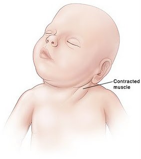
Appearance (see image): The head is tilted to one side (to the side of the affected muscle), and the chin is turned to the other side. There is stiffness, from the lack of movement, so there may be pain when the neck position is passively corrected.

A lump may be felt in the distal SCM.

Lump felt in distal SCM


 Investigation

The key is to differentiate between muscular torticollis (ie common, benign, easily correctible) and non-muscular torticollis (ie possibly secondary to neurological, ocular or vertebral pathology, and needing further investigation.

If there is a lump palpable in the SCM, it needs to be differentiated from other causes of a lump in the neck. Ultrasound is the best first line investigation – it detects fibrosis of the muscle (diagnosing torticollis) but would also pick up abnormal lymph nodes or masses.

Fine needle aspiration would be the next step if there was any uncertainty of the diagnosis, but this is rarely needed.

 Treatment

 Once muscular torticollis is confirmed:

 Physiotherapy is the mainstay of treatment. Even when there is severe fibrosis of the SCM, physio is effective in 98% [12]

  •  Neck stretches, performed regularly, moving the neck in the opposite direction to the affected muscle (tilt head sideways towards non-affected side, rotate towards affected side). Physio referral is indicated so parents can be taught the correct way to perform the stretches.
  • Let the baby spend more time lying on its tummy, to strengthen neck muscles
  • Use baby chair or Fraser chair to minimise the time the baby spends lying flat
  • Encourage head turning to affected side by using toys, distraction, feeding from that side
  • Physiotherapist may advise use of a neck brace in certain cases.

 (The above advice adapted from ‘Physio Questions’ [13], a blog by an Australian physio – torticollis featured as a blog entry in August 2010)


Surgical treatment is very rarely needed – only in instances where conservative management has failed after 6 months of treatment. When surgery is performed, the operation is a bipolar release of the SCM, and this has been found to be highly successful, even in patients older than 5 years [14] and into adulthood [15]

Alternatives to surgery?

A recent successful non-surgical development in treating cases resistant to physio is using botox injection. [16] The evidence for chiropractic treatment is weak, isolated successful cases have been described, [17] but there has been no randomised controlled trial. There are also reports of infants with torticollis caused by neurogenic tumours being treated (unsuccessfully) by a chiropractor before the correct diagnosis was made, [18] so it is imperative that parents have consulted a doctor before they choose to seek chiropractic help.

 References:

  1. http://www.ncbi.nlm.nih.gov/pubmed/3566514?tool=bestpractice.bmj.com
  2. http://www.ncbi.nlm.nih.gov/pubmed/21376202
  3. http://www.ncbi.nlm.nih.gov/pubmed/7484683
  4. http://www.ncbi.nlm.nih.gov/pubmed/18795328
  5. http://web.jbjs.org.uk/cgi/reprint/71-B/3/404
  6. http://www.ncbi.nlm.nih.gov/pubmed/21553338
  7. http://emedicine.medscape.com/article/1264848-overview
  8. http://www.ncbi.nlm.nih.gov/pubmed/22095422
  9. http://www.ncbi.nlm.nih.gov/pubmed/20638308
  10. http://www.ncbi.nlm.nih.gov/pubmed/8784707
  11. http://www.ncbi.nlm.nih.gov/pubmed/868283
  12. http://www.ncbi.nlm.nih.gov/pubmed/21843719
  13. http://physioquestions.blogspot.com/2010/08/are-you-worried-about-your-childs.html
  14. http://www.ncbi.nlm.nih.gov/pubmed/22045346
  15. http://www.ncbi.nlm.nih.gov/pubmed/19036153
  16. http://www.ncbi.nlm.nih.gov/pubmed/16470158
  17. http://www.ncbi.nlm.nih.gov/pubmed/8263436
  18. http://www.ncbi.nlm.nih.gov/pmc/articles/PMC2484567/?tool=pmcentrez

November’s Paediatric Pearls available now!

This is the first time in ages I’ve managed to get the finalised version on line in time for the end of the month!  NICE on autism this month, a bit on the use of corticosteroids in croup with help from the Cochrane Library, update on secondary prevention of meningococcal disease and a pointer to our local educational psychologist service which is hoping to make stronger links with the borough’s GPs (see also below).  Our feeding series continues with an article on colic from one of the junior paediatricians with lots of useful links and updates.